. 2017 Jun 23;17(1):148.
doi: 10.1186/s12862-017-0994-z.

Evolution of the vertebrate insulin receptor substrate (Irs) gene family

Affiliations

Evolution of the vertebrate insulin receptor substrate (Irs) gene family

Ahmad Al-Salam et al. BMC Evol Biol. .

Abstract

Background: Insulin receptor substrate (Irs) proteins are essential for insulin signaling as they allow downstream effectors to dock with, and be activated by, the insulin receptor. A family of four Irs proteins have been identified in mice, however the gene for one of these, IRS3, has been pseudogenized in humans. While it is known that the Irs gene family originated in vertebrates, it is not known when it originated and which members are most closely related to each other. A better understanding of the evolution of Irs genes and proteins should provide insight into the regulation of metabolism by insulin.

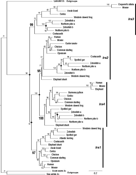
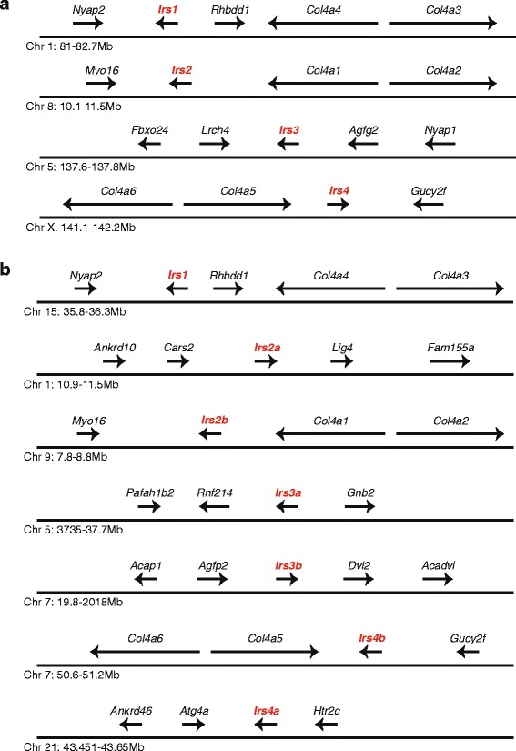
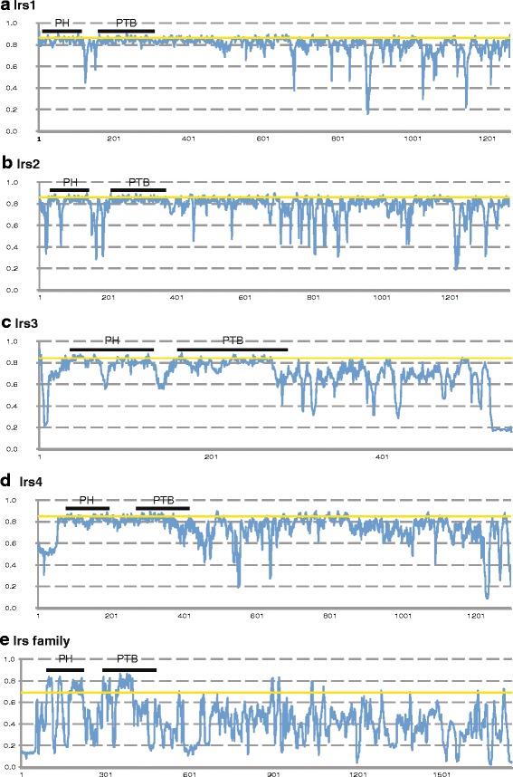
Results: Multiple genes for Irs proteins were identified in a wide variety of vertebrate species. Phylogenetic and genomic neighborhood analyses indicate that this gene family originated very early in vertebrae evolution. Most Irs genes were duplicated and retained in fish after the fish-specific genome duplication. Irs genes have been lost of various lineages, including Irs3 in primates and birds and Irs1 in most fish. Irs3 and Irs4 experienced an episode of more rapid protein sequence evolution on the ancestral mammalian lineage. Comparisons of the conservation of the proteins sequences among Irs paralogs show that domains involved in binding to the plasma membrane and insulin receptors are most strongly conserved, while divergence has occurred in sequences involved in interacting with downstream effector proteins.

Conclusions: The Irs gene family originated very early in vertebrate evolution, likely through genome duplications, and in parallel with duplications of other components of the insulin signaling pathway, including insulin and the insulin receptor. While the N-terminal sequences of these proteins are conserved among the paralogs, changes in the C-terminal sequences likely allowed changes in biological function.

Keywords: Episodic evolution; Gene duplication; Insulin receptor substrate; Phylogeny; Protein evolution; Pseudogene; Vertebrate.


Figures

Fig. 1
Fig. 1
Phylogeny of vertebrate Insulin receptor substrate (Irs) gene family sequences. Phylogeny of Irs sequences from diverse vertebrate species generated by Maximum likelihood using IQ-tree [49]. A similar phylogeny was generated by Bayesian methods [50, 51] (see Additional file 8: Figure S6). Phylogeny was rooted with sequences from acorn worm, purple sea urchin, and Florida lancelet (labeled as outgroups). Selected Irs sequences were chosen to represent the diversity of vertebrates, with different vertebrate Irs genes identified on the right. Similar results were obtained if other Irs sequences were used. Numbers at the nodes bootstrap support. Branch lengths are proportional to the inferred amount of change, with the scale bar at the bottom right. Diamonds indicate gene duplication events
Fig. 2
Fig. 2
Genomic organization of genes near Irs genes in the mouse and zebrafish genomes. The relative organization and orientation of genes near insulin receptor substrate (Irs) genes in (a) mouse and (b) zebrafish. Chromosomes and genomic locations are from Ensembl [44] (see Additional file 1: Table S1). Irs genes are labeled in red. Gene sizes and distances between genes are not to scale. Arrowheads indicate direction of transcription. Gene symbols are: Irs1–4, insulin receptors substrates 1–4; Col4a1–6, collagen, type IV, alpha1–6; Rhbdd1, rhomboid domain containing 1; Nyap2, Neuronal tyrosine-phophorylated phosphoinositide 3-kinase adaptor 2; Myo16, Myosin XVI; Fbxo24, F-box protein 24; Lrch4, Leucine-rich repeats and calponin homology (CH) domain containing 4; Agfp2, ArfGAP with FG repeats 2; Nyap1, Neuronal tyrosine-phosphorylated phosphoinositide 3-kinase adaptor 1; Gucy2f, Guanylate cyclase 2f; Ankrd10, Ankyrin repeat domain 10; Ankrd46, Ankyrin repeat domain 46; Cars2, Cysteinyl-tRNA synthetase 2; Lig4, Ligase IV, DNA, ATP-dependent; Fam155a, Family with sequence similarity 155, member A; Pafah1b2, Platelet-activating factor acetylhydrolase, isoform 1b, subunit 2; Rnf214, Ring finger protein 214; Gnb2, Guanine nucleotide binding protein (G protein), beta 2; Acap1, ArfGAP with coiled-coil, ankyrin repeat and PH domains 1; Dvl2, Dishevelled segment polarity protein 2; Acadvl, Acyl-Coenzyme A dehydrogenase, very long chain; Atg4a, Autophagy related 4A, cysteine peptidase; Htr2c, 5-hydroxytryptamine (serotonin) receptor 2C
Fig. 3
Fig. 3
Conservation of Irs protein sequences. JS divergence scores for aligned Irs protein sequences from 10 vertebrate species. (a) Irs1, (b) Irs2, (c) Irs3, (d) Irs4, (e) all Irs family members. JS scores are presented in Additional file 11: Table S4. Position in alignment is shown at the bottom of each graph. The locations of the PH and PTB are shown as bars near the top of each graph. JS scores above the yellow horizontal line are in the top 10% of JS scores for that alignment

References

    1. Pirola L, Johnston AM, Van Obberghen E. Modulation of insulin action. Diabetologia. 2004;47:170–184. doi: 10.1007/s00125-003-1313-3. - DOI - PubMed
    1. Myers MG, Jr, White MF. Insulin signal transduction and the IRS proteins. Annu Rev Pharmacol Toxicol. 1996;36:615–658. doi: 10.1146/annurev.pa.36.040196.003151. - DOI - PubMed
    1. De Meyts P, Whittaker J. Structural biology of insulin and IGF1 receptors: implications for drug design. Nat Rev Drug Discov. 2002;1:769–783. doi: 10.1038/nrd917. - DOI - PubMed
    1. Hubbard SR. The insulin receptor: both a prototypical and atypical receptor tyrosine kinase. Cold Spring Harb Perspect Biol. 2013;5:a008946. doi: 10.1101/cshperspect.a008946. - DOI - PMC - PubMed
    1. Ward CW, Lawrence MC. Ligand-induced activation of the insulin receptor: a multi-step process involving structural changes in both the ligand and the receptor. BioEssays. 2009;31:422–434. doi: 10.1002/bies.200800210. - DOI - PubMed

Substances

Grants and funding

LinkOut - more resources