. 2017 Jun 23;14(1):123.
doi: 10.1186/s12974-017-0900-z.

Identification of the flotillin-1/2 heterocomplex as a target of autoantibodies in bona fide multiple sclerosis

Affiliations

Identification of the flotillin-1/2 heterocomplex as a target of autoantibodies in bona fide multiple sclerosis

S Hahn et al. J Neuroinflammation. .

Abstract

Background: Autoantibodies, in particular those against aquaporin-4 and myelin-oligodendrocyte glycoprotein (MOG), aid as biomarkers in the differential diagnosis of demyelination. Here, we report on discovery of autoantibodies against flotillin in patients with multiple sclerosis (MS).

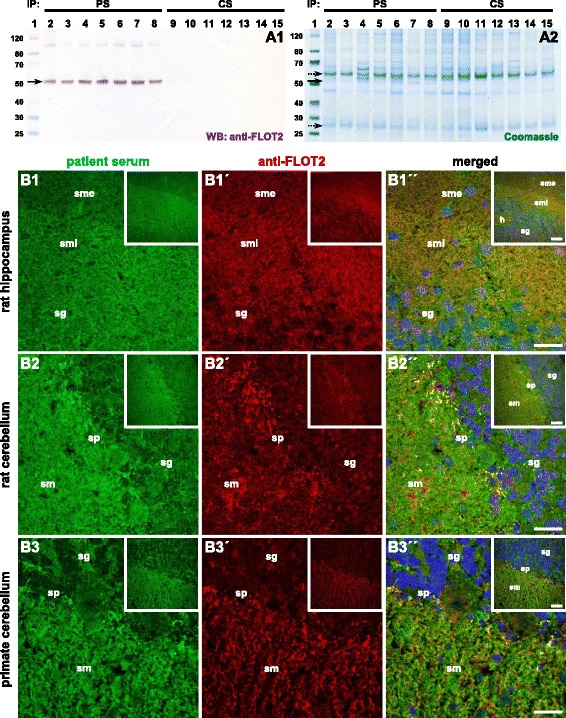
Methods: The target antigen was identified by histo-immunoprecipitation using the patients' sera and cryosections of rat or pig cerebellum combined with mass spectrometrical analysis. Correct identification was ascertained by indirect immunofluorescence and neutralization tests using the target antigens recombinantly expressed in HEK293 cells.

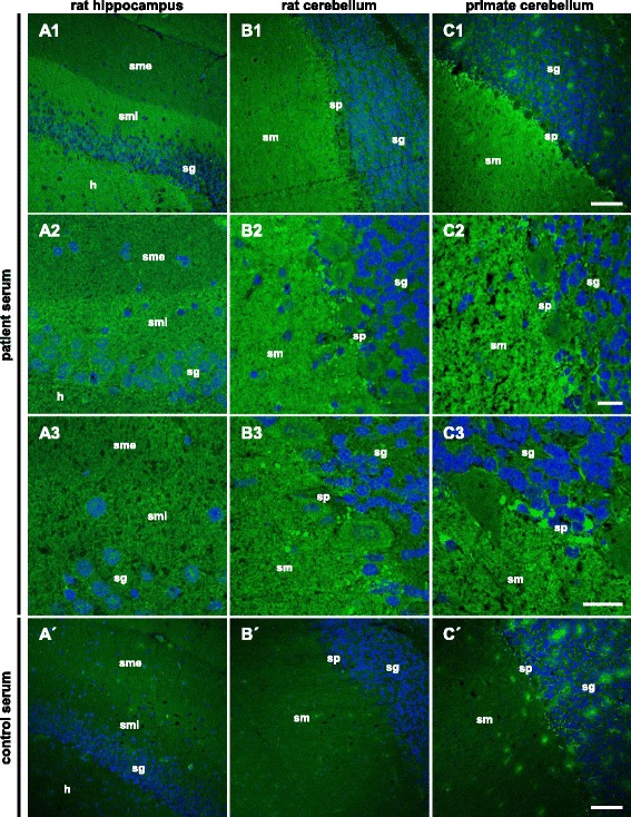
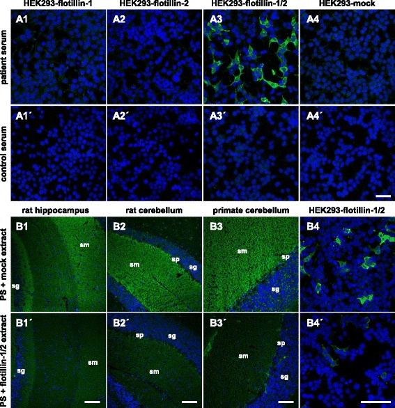
Results: Serum and CSF of the index patient produced a fine-granular IgG indirect immunofluorescence staining of the hippocampal and cerebellar molecular layers. Flotillin-1 and flotillin-2 were identified as target autoantigens. They also reacted with recombinant human flotillin-1/2 co-expressed in HEK293 cells, but not with the individual flotillins in fixed- and live-cell assays. Moreover, neutralization using flotillin-1/2, but not the single flotillins, abolished the tissue reactivity of patient serum. Screening of 521 patients, for whom anti-aquaporin-4 testing was requested and negative, revealed 8 additional patients with anti-flotillin-1/2 autoantibodies. All eight were negative for anti-MOG. Six patients ex post fulfilled the revised McDonald criteria for MS. Vice versa, screening of 538 MS sera revealed anti-flotillin-1/2 autoantibodies in eight patients. The autoantibodies were not found in a cohort of 67 patients with other neural autoantibody-associated syndromes and in 444 healthy blood donors.

Conclusions: Autoantibodies against the flotillin-1/2 heterocomplex, a peripheral membrane protein that is involved in axon outgrowth and regeneration of the optic nerve, are present in 1-2% of patients with bona fide MS.

Keywords: Autoantibodies; Flotillin; Histo-immunoprecipitation; Multiple sclerosis; Optic neuritis; Reggie.


Figures

Fig. 1
Fig. 1
Immunofluorescence staining of central nervous tissues. Cryosections of rat hippocampus (A) and cerebellum (B) as well as primate cerebellum (C) were incubated with patient serum (AC) or with control serum (A′–C′) (AC1, A′–C′ 1:100, AC2, C3 1:50) in the first step and with Alexa Fluor 488-labeled goat anti-human IgG in the second step (green). Nuclei were counterstained by incubation with TO-PRO-3 iodide (blue). A fine-granular staining of the stratum moleculare (sm) was obtained. On the hippocampus, the sm internum was stained more intensely than the sm externum. Scale bar: 100 μm (AC1, A′–C′), 20 μm (AC2, C3). h hilus, sm stratum moleculare, smi stratum moleculare internum, sme stratum moleculare externum, sg stratum granulosum, sp stratum purkinjense
Fig. 2
Fig. 2
Histo-immunoprecipitation and antigen identification. Cryosections of rat or pig cerebellum were incubated with the serum (1:100), washed in PBS, and solubilized using detergents. The solution was incubated with protein-G-coated magnetic beads. The immunocomplexes were eluted by SDS and subjected to SDS-PAGE analysis and Western blot. A Western blot after incubation with anti-flotillin-2 (A1) and enzymatic visualization of antibody binding. Staining of SDS polyacrylamide gel with colloidal Coomassie (A2). Lane 1: molecular mass (kDa) marker; lanes 2–8: histo-immunoprecipitates of patient sera from rat cerebellum; lanes 9–15: histo-immunoprecipitates of control samples. The arrow indicates the position of the immunoprecipitated antigen 50 kDa while dotted arrows indicate the position of IgG heavy and light chain at 52 and 27 kDa, respectively. PS patient sample, CS control sample. B Immunofluorescence staining of rat hippocampus (B1) and cerebellum (B2) and primate cerebellum (B3) tissue sections with patient serum (green, 1–3) and anti-flotillin-2 antibody (red, 1′–3′). The merged images show localization of the reactivity in the same region including the more intense staining of the sm internum on the hippocampus (1″–3″). Scale bar: 50 μm (large images), 100 μm (inserts). PS patient serum, CS control serum, h hilus, sg stratum granulosum, sm stratum moleculare, smi stratum moleculare internum, sme stratum moleculare externum, sp stratum purkinjense
Fig. 3
Fig. 3
Immunofluorescence staining of recombinant flotillin and the neutralization of antibody reaction on tissue. A Immunofluorescence analysis of transfected HEK293 cells. Acetone-fixed recombinant HEK293 cells expressing flotillin-1 (1), flotillin-2 (2), flotillin-1 and flotillin-2 (3) or a mock-transfected control (4) were incubated with patient serum (1–4) or with control serum (1′–4′) (both 1:100). Cell nuclei were counterstained with TO-PRO-3 iodide (blue). Only HEK293-flotillin-1/2 cells reacted with the samples (green). Scale bar: 50 μm. B Neutralization of immunofluorescence reaction on neuronal tissues. Serum was pre-incubated with extracts of HEK293 cells transfected with empty control vector (1–4) or with the plasmid harboring flotillin-1/2 cDNA (1′–4′). The extract containing flotillin-1/2 greatly reduced or abolished the immune reaction of the serum on rat hippocampus (1′) and cerebellum (2′), primate cerebellum (3′), and HEK293-flotillin-1/2 (4′). The control extracts had no effect (1–4). Nuclei were counterstained by incubation with TO-PRO-3 iodide (blue). Scale bar: 50 μm. PS patient serum, CS control serum, sg stratum granulosum, sm stratum moleculare, sp stratum purkinjense

References

    1. Probst C, Saschenbrecker S, Stöcker W, Komorowski L. Anti-neuronal autoantibodies: current diagnostic challenges. Mult Scler Rel Dis. 2014;3:304–320. - PubMed
    1. Lennon VA, Kryzer TJ, Pittock SJ, Verkman AS, Hinson SR. IgG marker of optic-spinal multiple sclerosis binds to the aquaporin-4 water channel. J Exp Med. 2005;202:473–477. doi: 10.1084/jem.20050304. - DOI - PMC - PubMed
    1. Dalmau J, Gleichman AJ, Hughes EG, Rossi JE, Peng X, Lai M, Dessain SK, Rosenfeld MR, Balice-Gordon R, Lynch DR. Anti-NMDA-receptor encephalitis: case series and analysis of the effects of antibodies. Lancet Neurol. 2008. - PMC - PubMed
    1. Lancaster E, Lai M, Peng X, Hughes E, Constantinescu R, Raizer J, Friedman D, Skeen MB, Grisold W, Kimura A, Ohta K, Iizuka T, Guzman M, Graus F, Moss SJ, Balice-Gordon R, Dalmau J. Antibodies to the GABA(B) receptor in limbic encephalitis with seizures: case series and characterisation of the antigen. Lancet Neurol. 2010;9(1):67-76. - PMC - PubMed
    1. Irani SR, Alexander S, Waters P, Kleopa KA, Pettingill P, Zuliani L, Peles E, Buckley C, Lang B, Vincent A. Antibodies to Kv1 potassium channel-complex proteins leucine-rich, glioma inactivated 1 protein and contactin-associated protein-2 in limbic encephalitis, Morvan’s syndrome and acquired neuromyotonia. Brain. 2010;133:2734–2748. doi: 10.1093/brain/awq213. - DOI - PMC - PubMed

Publication types