. 2017 Jun 23;14(1):21.
doi: 10.1186/s12989-017-0202-8.

MyD88-dependent pro-interleukin-1β induction in dendritic cells exposed to food-grade synthetic amorphous silica

Affiliations

MyD88-dependent pro-interleukin-1β induction in dendritic cells exposed to food-grade synthetic amorphous silica

Hans Christian Winkler et al. Part Fibre Toxicol. .

Abstract

Background: Dendritic cells (DCs) are specialized first-line sensors of foreign materials invading the organism. These sentinel cells rely on pattern recognition receptors such as Nod-like or Toll-like receptors (TLRs) to launch immune reactions against pathogens, but also to mediate tolerance to self-antigens and, in the intestinal milieu, to nutrients and commensals. Since inappropriate DC activation contributes to inflammatory diseases and immunopathologies, a key question in the evaluation of orally ingested nanomaterials is whether their contact with DCs in the intestinal mucosa disrupts this delicate homeostatic balance between pathogen defense and tolerance. Here, we generated steady-state DCs by incubating hematopoietic progenitors with feline McDonough sarcoma-like tyrosine kinase 3 ligand (Flt3L) and used the resulting immature DCs to test potential biological responses against food-grade synthetic amorphous silica (SAS) representing a common nanomaterial generally thought to be safe.

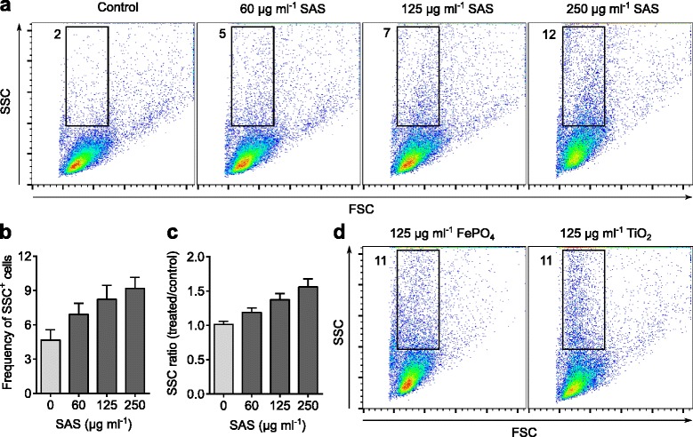
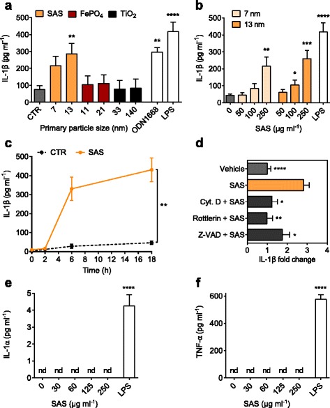
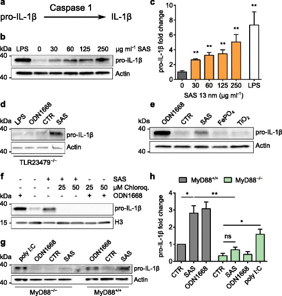
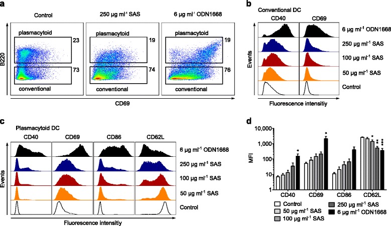
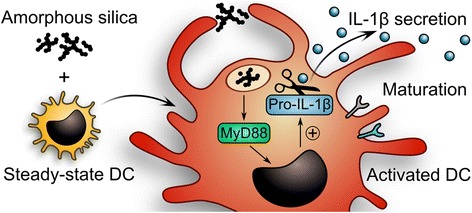
Results: Interaction of immature and unprimed DCs with food-grade SAS particles and their internalization by endocytic uptake fails to elicit cytotoxicity and the release of interleukin (IL)-1α or tumor necrosis factor-α, which were identified as master regulators of acute inflammation in lung-related studies. However, the display of maturation markers on the cell surface shows that SAS particles activate completely immature DCs. Also, the endocytic uptake of SAS particles into these steady-state DCs leads to induction of the pro-IL-1β precursor, subsequently cleaved by the inflammasome to secrete mature IL-1β. In contrast, neither pro-IL-1β induction nor mature IL-1β secretion occurs upon internalization of TiO2 or FePO4 nanoparticles. The pro-IL-1β induction is suppressed by pharmacologic inhibitors of endosomal TLR activation or by genetic ablation of MyD88, a downstream adapter of TLR pathways, indicating that endosomal pattern recognition is responsible for the observed cytokine response to food-grade SAS particles.

Conclusions: Our results unexpectedly show that food-grade SAS particles are able to directly initiate the endosomal MyD88-dependent pathogen pattern recognition and signaling pathway in steady-state DCs. The ensuing activation of immature DCs with de novo induction of pro-IL-1β implies that the currently massive use of SAS particles as food additive should be reconsidered.

Keywords: E 551; Food additive; Food toxicology; Gut-associated lymphoid tissue; Inflammatory bowel disease; Nanomaterial; Silicon dioxide; Synthetic amorphous silica.


Figures

Fig. 1
Fig. 1
Interaction of steady-state DCs with nanomaterials. Flt3L-generated immature DCs were incubated for 1 h at 37 °C with the indicated concentrations of SAS (13-nm primary diameter), 11-nm FePO4 or 33-nm TiO2 particles, and analyzed by flow cytometry. In culture medium, the SAS particles form aggregates with a mean diameter of 127 nm. The forward scatter (FSC) depends on cell size whereas the side scatter (SSC) reflects intracellular contents like granules [34, 35]. a Flow cytometry distributions demonstrating a SAS dose-dependent increase of DCs with elevated SSC values (numbers denote percentages of events in each gate). b Mean percentage of cells in the selected gate (shown in a) with high SSC values. Upon one-way ANOVA, SAS treatments increased the proportion of high-SSC cells in a significant manner (p < 0.05, n = 4 experiments with independent bone marrow isolates). Error bars, standard errors of the mean (s.e.m.). c Ratios of median SSC. Upon one-way ANOVA, SSC values after incubation with SAS particles were significantly higher than controls (p < 0.05, n = 4). d Comparison with SSC increments resulting from incubation of DCs with FePO4 and TiO2 nanoparticles (quantifications are shown in Additional file 1: Figure S3)
Fig. 2
Fig. 2
Internalization of nanomaterials by steady-state DCs. Immature DCs were incubated for 2 h at 37 °C with 250 μg ml−1 SAS particles (13-nm primary diameter) and analyzed by TEM. a Representative steady-state DC showing emerging dendrites interacting with particles. N, nucleus; bar, 0.5 μm. Contrasted with uranyl acetate/lead citrate for 15 min; the rectangle indicates the area selected for higher magnification. b Magnified region of the DC near its cell surface to highlight membrane protrusions (arrowhead) in the process of engulfing SAS particles (asterisk). The two arrows indicate internalized particles within a vacuole (V). Scale bar, 0.2 μm. c Magnified region of a DC that visualizes the process by which SAS particles (asterisk) are engulfed into intracellular vacuoles. A small SAS aggregate is enclosed in a vacuole. Scale bar, 0.2 μm; contrasted with uranyl acetate/lead citrate for 1 min to improve particle visibility. d Analysis of a representative intracellular SAS particle aggregate and cytoplasmic background by energy-dispersive X-ray spectroscopy (EDX). Internalized FePO4 and TiO2 particles are shown in Additional file 1: Figure S5
Fig. 3
Fig. 3
IL-1β secretion induced by food-grade SAS particles depends on uptake by macropinocytosis. Immature DCs were incubated (18 h, 37 °C) with particles to test for IL-1β secretion. Asterisks denote significant differences between SAS treatments and controls (*p < 0.05, **p < 0.01, ***p < 0.001, ****p < 0.0001). a DCs were exposed to particles (250 μg ml−1) and supernatants analyzed for IL-1β. Control reactions contained medium (CTR), 6 μg ml−1 ODN1668 (mimicking microbial DNA) or 1 μg ml−1 lipopolysaccharide (LPS). One-way ANOVA with Dunnet’s correction; n = 3–9; error bars, s.e.m. b Dose dependence of IL-1β secretion stimulated by SAS particles. Unpaired two-tailed t-test (n = 3–12). c Time dependence of IL-1β secretion stimulated by SAS particles (250 μg ml−1). Unpaired two-tailed t-test (n = 4). d Flt3L-generated immature DCs were incubated for 18 h at 37 °C with 13-nm SAS (250 μg ml−1) alone or in the presence of cytochalasin D (1.5 μg ml−1), rottlerin (1.5 μg ml−1) or Z-VAD (10 μg ml−1). Results represent fold changes relative to vehicle controls (one-way ANOVA with Dunnet’s correction, n = 3–12). e Release of IL-1α into the cell culture supernatant stimulated by LPS (1 μg ml−1) but not by 13-nm SAS particles. Asterisks denote significant differences between the LPS treatment and controls containing only culture medium. Cytokine levels below detection limit (4 pg ml−1) are indicated as not detectable (nd). One-way ANOVA with Dunnet’s correction; n = 4; error bars, s.e.m. f Release of TNF-α into the cell culture supernatant stimulated by LPS (1 μg ml−1) but not by 13-nm SAS particles (nd, cytokine levels below the detection limit of 8 pg ml−1. Asterisks denote significant differences between the LPS treatment and controls containing only culture medium. One-way ANOVA with Dunnet’s correction; n = 4; error bars, s.e.m.
Fig. 4
Fig. 4
Induction of pro-IL-1β by food-grade SAS particles depends on MyD88. Immature DCs were incubated (18 h, 37 °C) with particles to test for IL-1β induction. Asterisks denote significant differences between SAS treatments and controls (*p < 0.05, **p < 0.01, ***p < 0.001, ****p < 0.0001). a Schematic illustrating the mechanism of IL-1β production. b DCs were incubated with LPS (250 ng ml−1) or 13-nm SAS and analyzed for pro-IL-1β (31 kDa) and actin (42 kDa) by immunoblotting. c Quantification of pro-IL-1β induction by SAS particles (unpaired two-tailed t-test, n = 5, error bars, s.e.m.). d Incubation of TLR2/3/4/7/9−/− DCs with LPS (250 ng ml−1), ODN1668 (600 ng ml−1), medium (CTR) or SAS particles (125 μg ml−1). e Incubation of wild type DCs with ODN1668 (600 ng ml−1), medium or the indicated particles (125 μg ml−1). f Effect of endosomal TLR inhibition. DCs were incubated with 13-nm SAS (125 μg ml−1) alone or in the presence of chloroquine and analyzed for pro-IL-1β and histone H3 (17 kDa) by immunoblotting. ODN1668 (600 ng ml−1) served as positive control. g MyD88−/− or wild type (MyD88+/+) DCs were incubated with poly I:C (5 μg ml−1), ODN1668 (600 ng ml−1), medium (CTR) or SAS particles (125 μg ml−1) and analyzed for pro-IL-1β (31 kDa) and actin (42 kDa) by immunoblotting. Split bands in some control lanes are an electrophoretic artifact not interfering with quantifications. h Pro-IL-1β induction by SAS particles in MyD88−/− or wild type (MyD88+/+) DCs (unpaired two-tailed t-test, n = 3, error bars, s.e.m.)
Fig. 5
Fig. 5
Maturation markers on steady-state DCs. Immature DCs were incubated for 18 h at 37 °C with the indicated stimulus and their surface markers were analyzed by flow cytometry. a Increased CD69 on the surface of DCs exposed to SAS particles (13-nm primary diameter) or oligonucleotide ODN1668. Plasmacytoid and conventional DCs are the two major subsets differing in B220 expression (numbers denote percentages of events in each gate). b Representative histograms showing dose-dependent changes of CD69 and CD40 on conventional DCs exposed to the indicated stimuli. Control, unstimulated DCs in culture medium. c Representative histograms showing dose-dependent changes in the display by CD69, CD40, CD86 and CD62L on plasmacytoid DCs. d Quantification of maturation markers by median fluorescence intensity (MFI) on plasmacytoid DCs exposed to SAS particles (13-nm primary size). Statistical significance (*p < 0.05, **p < 0.01, ***p < 0.001) was determined by one-way ANOVA with Dunnet’s correction, n = 3 experiments with independent bone marrow isolates; error bars, s.e.m
Fig. 6
Fig. 6
Scheme summarizing the single-hit mechanism of IL-1β secretion by steady-state DCs exposed to food-grade SAS particles. In immature DCs, pro-IL-1β is not expressed, but its induction takes place upon uptake of SAS particles into endosomes and MyD88-dependent TLR signaling. Cleavage of the newly expressed pro-IL-1β precursor by the inflammasome-associated caspase leads to IL-1β secretion. In addition, the immature DCs undergo maturation involving shifts in multiple surface markers. IL-1β secretion in response to SAS particles is suppressed by cytochalasin or rottlerin (inhibitors of actin-dependent macropinocytosis), by chloroquine or bafilomycin A (inhibitors of endosomal TLR activation), by genetic ablation of MyD88 (the central adapter of TLR signaling) and by Z-VAD (an inhibitor of the inflammasome-associated caspase)

Similar articles

Cited by

References

    1. Watowich SS, Liu Y-J. Mechanisms regulating dendritic cell specification and development. Immunol Rev. 2010;238:76–92. doi: 10.1111/j.1600-065X.2010.00949.x. - DOI - PMC - PubMed
    1. Shortman K, Naik SH. Steady-state and inflammatory dendritic-cell development. Nat Rev Immunol England. 2007;7:19–30. doi: 10.1038/nri1996. - DOI - PubMed
    1. Bogunovic M, Ginhoux F, Helft J, Shang L, Hashimoto D, Greter M, et al. Origin of the lamina propria dendritic cell network. Immunity. 2009;31:513–525. doi: 10.1016/j.immuni.2009.08.010. - DOI - PMC - PubMed
    1. Varol C, Vallon-Eberhard A, Elinav E, Aychek T, Shapira Y, Luche H, et al. Intestinal lamina propria dendritic cell subsets have different origin and functions. Immunity. 2009;31:502–512. doi: 10.1016/j.immuni.2009.06.025. - DOI - PubMed
    1. Pandey S, Kawai T, Akira S. Microbial sensing by Toll-like receptors and intracellular nucleic acid sensors. Cold Spring Harb Perspect Biol. 2015;7:a016246. doi: 10.1101/cshperspect.a016246. - DOI - PMC - PubMed

Publication types

MeSH terms

LinkOut - more resources