. 2017:591:355-414.
doi: 10.1016/bs.mie.2017.03.024. Epub 2017 May 10.

Electrical Probes of DNA-Binding Proteins

Affiliations

Electrical Probes of DNA-Binding Proteins

Jacqueline K Barton et al. Methods Enzymol. 2017.

Abstract

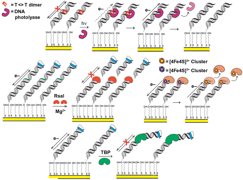
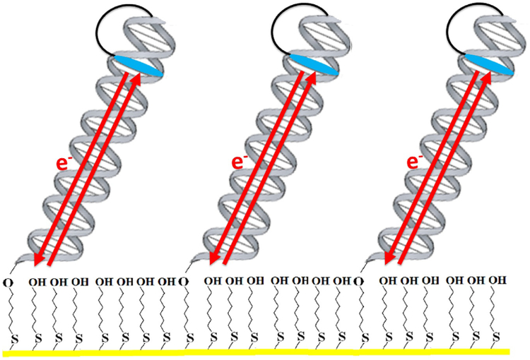
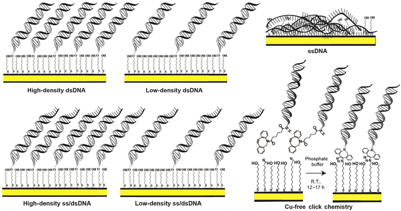
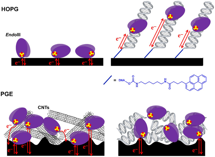
A DNA electrochemistry platform has been developed to probe proteins bound to DNA electrically. Here gold electrodes are modified with thiol-modified DNA, and DNA charge transport chemistry is used to probe DNA binding and enzymatic reaction both with redox-silent and redox-active proteins. For redox-active proteins, the electrochemistry permits the determination of redox potentials in the DNA-bound form, where comparisons to DNA-free potentials can be made using graphite electrodes without DNA modification. Importantly, electrochemistry on the DNA-modified electrodes facilitates reaction under aqueous, physiological conditions with a sensitive electrical measurement of binding and activity.

Keywords: Base excision repair; Base flipping; DNA alkylation repair; DNA binding; DNA glycosylase; DNA modification; Kinetic simulation; Nucleotide flipping; Stopped flow; Transient kinetics.


Figures

Fig. 1
Fig. 1
Electrochemical monitoring of DNA-binding protein activity on DNA-modified electrodes. (Top) DNA photolyase binds and repairs a thymine–thymine dimer on a DNA-modified electrode, restoring DNA CT and producing a signal from the flavin cofactor, through repaired DNA (DeRosa, Sancar, & Barton, 2005). (Center left) RsaI restriction enzyme cuts duplex DNA, removing covalently attached redox probe. Signal disappears after wash of surface, indicating that RsaI binding and cutting of DNA at recognition site occurs (Slinker, Muren, Renfrew, & Barton, 2011). (Center right) A bound [4Fe4S] enzyme is oxidized from the resting [4Fe4S]2+ state to the tightly bound [4Fe4S]3+ state through DNA CT; it can then be reduced from the tightly bound [4Fe4S]3+ state to the more weakly binding resting [4Fe4S]2+ state through DNA CT, promoting dissociation (Boal et al., 2009). (Bottom) TBP binding kinks duplex DNA, attenuating CT, and diminishing signal from a DNA-intercalating, covalently attached redox probe (Gorodetsky, Ebrahim, & Barton, 2008).
Fig. 2
Fig. 2
Electrochemical monitoring of DNA-mediated charge transport processes. In a typical setup, alkanethiol-modified DNA is annealed to its complement and allowed to form a self-assembled monolayer on a gold electrode. Gaps in the Au surface are filled in with 6-mercapto-1-hexanol, passivating the surface, and electrochemistry is carried out in a buffered solution. Redox-active probes, such as the intercalator Nile Blue, can be covalently tethered to one end of the DNA, or simply bound noncovalently. The DNA duplex then serves as a bridge for electron transfer between the probe and the gold electrode. Notably, charge transport through the DNA is very rapid, and electron transfer rates in this system are limited by tunneling through the alkanethiol linker.
Fig. 3
Fig. 3
Different DNA monolayer morphologies formed on DNA-modified Au electrodes. When duplex DNA is incubated with Mg2+ on an Au surface (yellow), the substrate forms a high-density monolayer of duplex DNA (top left). When incubated on Au in the absence of Mg2+ a low-density duplex DNA monolayer results. DNA containing a single-stranded overhang segment at the interface of DNA monolayer and electrolyte can also be used to form high-density or low-density monolayers for assaying proteins with a preferred primed end substrate (bottom left). When single-stranded DNA is incubated on the Au electrode, the substrate adheres to the surface and passivates the Au, precluding observation of a redox signal (top right). Finally, Cu-free click chemistry can be used to form a DNA monolayer on an Au electrode surface (bottom right). Azide-terminated alkanethiol-modified Au electrode is incubated in 1:1 mix of mercaptoundecanol and 1-azidoundecane-11-thiol in ethanol for about 4h. 50 μM DBCO-modified dsDNA in DNA phosphate buffer is incubated with modified Au electrodes for 12–17h to let the cyclooctyne-based copper-free click reaction proceed. DBCO-modified DNA clicks only to the azide terminal groups, so that the binding density depends on the initial azide content. These monolayers all serve as useful conditions or controls when characterizing redox activity of a DNA-binding enzyme.
Fig. 4
Fig. 4
Different platforms for DNA electrochemistry. Single Au electrodes can be set up on either an Au on mica surface (left) or using a rod electrode (right). A multiplex platform (center) (Pheeney et al., 2013; Slinker et al., 2010) with 16-electrodes separated into four quadrants can also be used to assay multiple DNA substrates on a single surface, with replicates for each condition. Platforms are shown from the top (above) and from the side (below) with components of the setup.
Fig. 5
Fig. 5
Electrocatalytic cycle between free methylene blue (MB) and ferricyanide on a DNA-modified electrode. MB in its oxidized form is intercalated into the DNA base stack. Upon reduction of MB to leucomethylene blue (LB) via DNA-mediated CT, the affinity of the LB for DNA is lowered, and LB is no longer intercalated. The reduced LB is capable of reducing ferricyanide that is freely diffusing in solution. The LB is then reoxidized to MB and can reintercalate into the DNA. The ferricyanide acts as a diffusing electron sink in solution for the redox probe MB. Electrostatic repulsion prevents ferricyanide from penetrating the negatively charged DNA film. A cyclic voltammetry at a DNA-modified electrode of ferricyanide (black), MB (blue), and ferricyanide and MB (red).
Fig. 6
Fig. 6
Overview of electrochemical DNMT1 analysis from tumors with two-electrode platform (top). Tumor and healthy tissues are lysed, and nuclear lysate is used to detect DNMT1 methyltransferase activity. The lysate is applied to a multiplexed, two working electrode platform that enables the conversion of methylation events into an electro-chemical signal. The electrochemical detection platform contains two electrode arrays, each with 15 electrodes (1mm diameter each) in a 5 × 3 array. Multiple DNAs are patterned covalently to the substrate electrode by an electrochemically activated click reaction initiated with the patterning electrode array. Once a DNA array is established on the substrate electrode platform, electrocatalytic detection is then performed from the top patterning/detection electrode. Generally, we find hyperactivity of DNMT1 in tumor samples as compared to the healthy adjacent tissue. Signal-on electrochemical assay for DNMT1 detection (bottom). Left: The bottom (primary) electrode modified with a dilute DNA monolayer is responsible for generating electrochemical signals through DNA-mediated (CT) amplified by electrocatalysis. Methylene blue (MB), a DNA-intercalating redox probe, is reduced by DNA CT and enters solution as leucomethylene blue (LB), where it can interact with an electron sink, ferricyanide. Upon interaction with LB, ferricyanide is reduced to ferrocyanide, reoxidizing the LB to MB in the process. Current is generated and detected at the secondary electrode from the reoxidation of ferrocyanide. The current generated is proportional to the amount of ferrocyanide oxidized. To detect DNMT1, crude lysate is added to the electrode. If DNMT1 (blue) is capable of methylating DNA (red arrow), the DNA on the electrode becomes fully methylated. If the protein is not active, the DNA remains hemimethylated or unmethylated (green arrow). A methylation-specific restriction enzyme BssHII (purple) is then added that cuts the unmethylated or hemimethylated DNA (green arrow), significantly attenuating the electrochemical signal, while leaving the fully methylated DNA (red arrow) untouched. Constant potential amperometry (right) is used to measure the percent change before and after restriction enzyme treatment. If the restriction enzyme does not affect the DNA (top), the signals overlay. If, however, the restriction enzyme cuts the DNA, the signal is significantly attenuated (bottom).
Fig. 7
Fig. 7
Electrochemistry of EndoIII and MutY on DNA-modified gold electrodes. EndoIII and MutY are BER glycosylases that target sites of oxidative damage in DNA; EndoIII (top left) excises oxidized pyrimidines, while MutY (top right) removes adenine mispaired with 8oxoG. When incubated on a DNA-modified electrode, both proteins (EndoIII depicted) display reversible single-electron redox peaks by CV, a process that can be disrupted by mutating critical amino acid residues in the CT pathway as illustrated by EndoIII Y82A (bottom). Structures are adapted from PDB structures IP59 (EndoIII) and 1RRQ (MutY); both are from Geobacillus stearothermophilus, but each shows high homology to the E. coli proteins used in electrochemistry. The CV is adapted from Pheeney, C. G., Arnold, A. R., Grodick, M. A., & Barton, J. K. (2013). Multiplexed electrochemistry of DNA-bound metalloproteins. Journal of the American Chemical Society, 135, 11869–11878.
Fig. 8
Fig. 8
Electrochemistry of the NER helicase XPD on a substrate containing a 9-mer 5’ single-stranded overhang. On this substrate, electro-chemical experiments with XPD yielded a signal similar in potential and general form to those from BER proteins (left, blue CV at right). The addition of ATP, known to stimulate helicase activity in XPD, resulted in a substantial increase in current as a result of enhanced electronic coupling between the [4Fe4S] cluster and the DNA base stack (middle, red CV at right). All images in this figure were adapted from Mui, T. P., Fuss, J. O., Ishida, J. P., Tainer, J. A., & Barton, J. K. (2011) ATP-stimulated, DNA-mediated redox signaling by XPD, a DNA repair and transcription helicase. Journal of the American Chemical Society, 133, 16378–16381.
Fig. 9
Fig. 9
Graphite platforms for protein electrochemistry. Two general platforms are commonly used for protein electrochemistry: HOPG (top) and PGE (bottom). HOPG consists of a pristinely flat, strongly hydrophobic surface, while PGE is rough and often contains surface oxides that lower the hydrophobicity. Proteins can adsorb directly to HOPG, although this interaction is weak, but the ability of pyrene-modified DNA to form a noncovalent bond with the surface allows a direct comparison of DNA-free and DNA-bound proteins. In contrast, PGE provides ample surface area for binding DNA-free proteins, and signals can be further enhanced by the addition of carbon nanotubes (CNTs); DNA can be incorporated into a film on PGE, but in this environment, it tends to passivate the surface and the random orientation prevents the observation of DNA-mediated signals.

References

    1. Agard NJ, Prescher JA, & Bertozzi CR (2004). A strain-promoted [3+2] azidealkyne cycloaddition for covalent modification of blomolecules in living systems. Journal of the American Chemical Society, 126, 15046–15047. - PubMed
    1. An R, Jia Y, Wan B, Zhang Y, Dong P, Li J, et al. (2014). Non-enzymatic depurination of nucleic acids: Factors and mechanisms. PloS One, 9, e115950. - PMC - PubMed
    1. Armstrong FA, Bond AM, Hill HAO, Oliver N, & Psalti ISM (1989). Electrochemistry of cytochrome c, plastocyanin, and ferredoxin at edge- and basal-plane graphite electrodes interpreted via a model based on electron transfer at electroactive sites of microscopic dimensions in size. Journal of the American Chemical Society, 111, 9185–9189.
    1. Arnold AR, Grodick MA, & Barton JK (2016). DNA charge transport: From chemical principles to the cell. Cell Chemical Biology, 23, 183–197. - PMC - PubMed
    1. Baffert C, Sybirna K, Ezanno P, Lautier T, Hajj V, Meynial-Salles I, et al. (2012). Covalent attachment of FeFe hydrogenases to carbon electrodes for direct electron transfer. Analytical Chemistry, 84, 7999–8005. - PubMed

Publication types

LinkOut - more resources