. 2017 Jul:21:170-181.
doi: 10.1016/j.ebiom.2017.06.011. Epub 2017 Jun 13.

Inhibition of Non Canonical HIV-1 Tat Secretion Through the Cellular Na+,K+-ATPase Blocks HIV-1 Infection

Affiliations

Inhibition of Non Canonical HIV-1 Tat Secretion Through the Cellular Na+,K+-ATPase Blocks HIV-1 Infection

Silvia Agostini et al. EBioMedicine. 2017 Jul.

Abstract

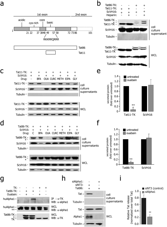
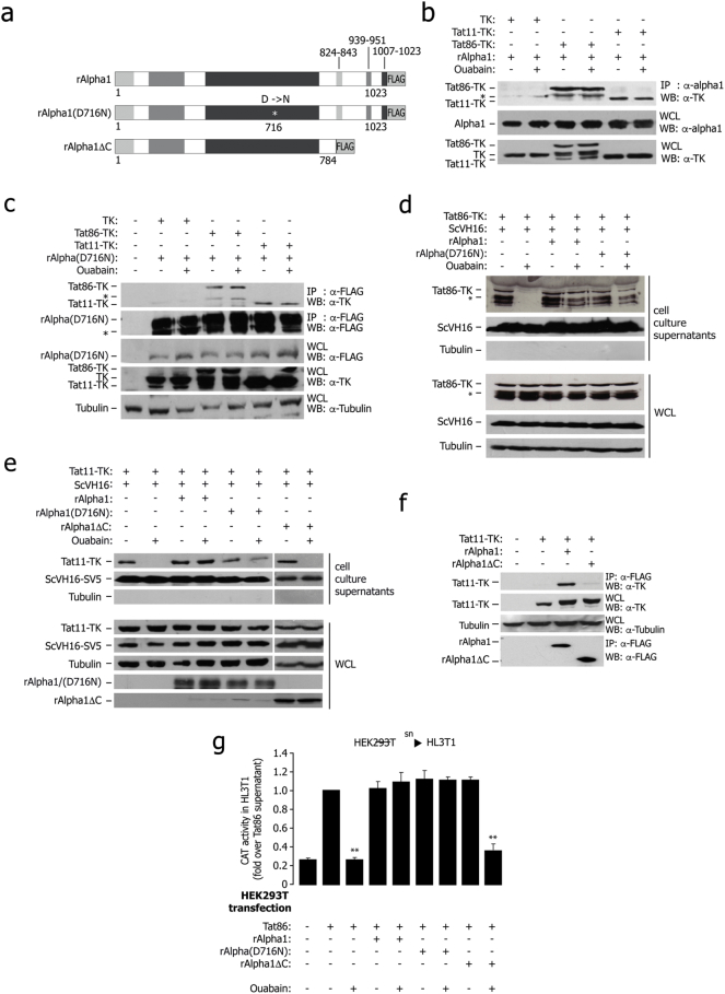
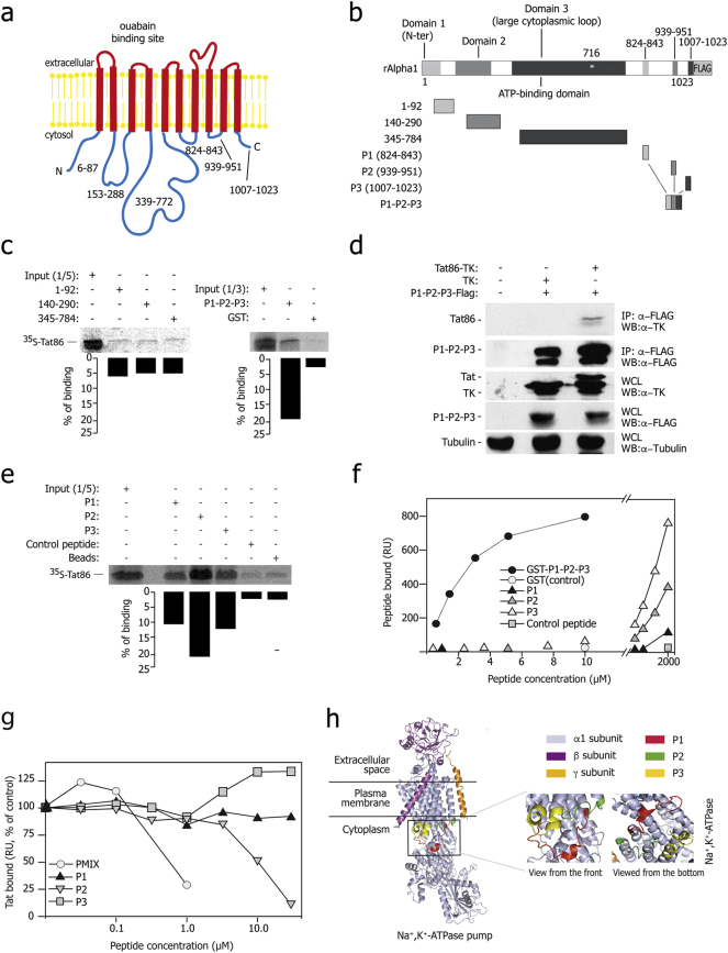
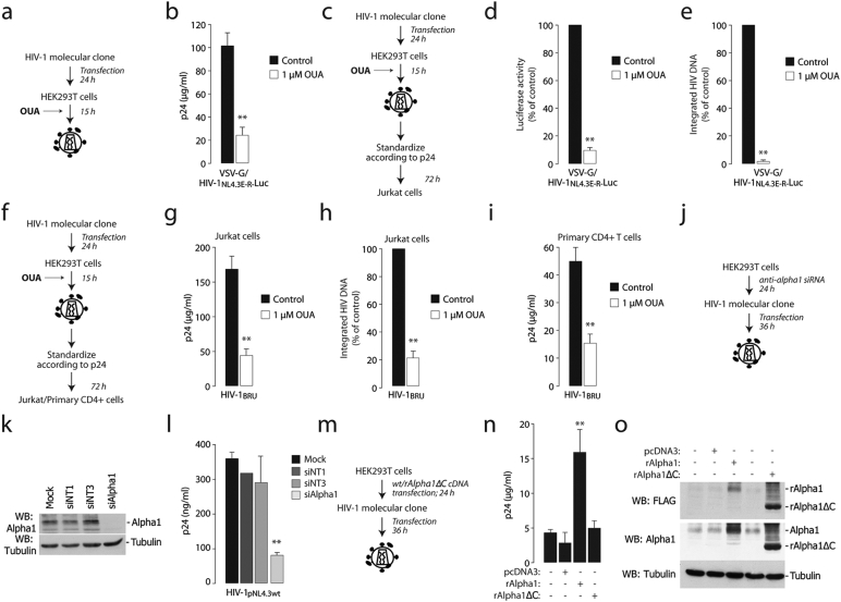
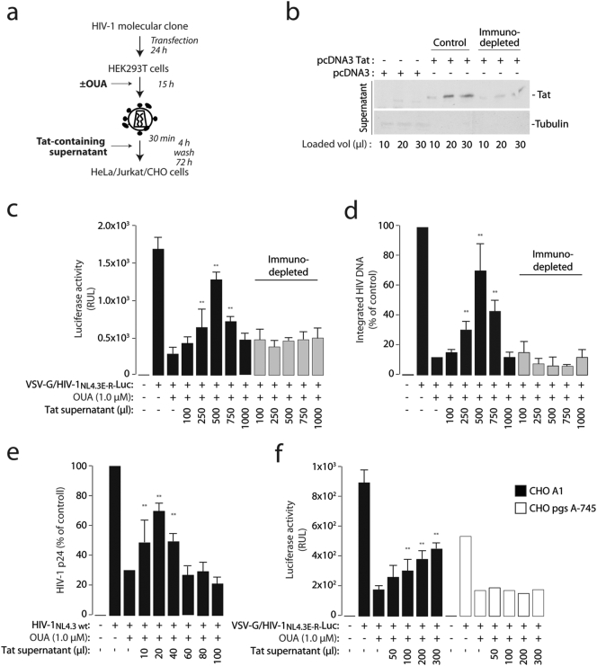
Besides its essential role in the activation of HIV-1 gene expression, the viral Tat protein has the unusual property of trafficking in and out of cells. In contrast to Tat internalization, the mechanism involved in extracellular Tat release has so far remained elusive. Here we show that Tat secretion occurs through a Golgi-independent pathway requiring binding of Tat with three short, non-consecutive intracytoplasmic loops at the C-terminus of the cellular Na+,K+-ATPase pump alpha subunit. Ouabain, a pump inhibitor, blocked this interaction and prevented Tat secretion; virions produced in the presence of this drug were less infectious, consistent the capacity of virion-associated Tat to increase HIV-1 infectivity. Treatment of CD4+ T-cells with short peptides corresponding to the Tat-binding regions of the pump alpha subunit impaired extracellular Tat release and blocked HIV-1 replication. Thus, non canonical, extracellular Tat secretion is essential for viral infectivity.

Keywords: ATPase; HIV-1; Protein secretion; Surface plasmon resonance; Tat; Transactivation.


Figures

Fig. 1
Fig. 1
Ouabain-sensitive secretion of Tat from the expressing cells. (a) Schematic representation of the major functional domains of HIV-1 Tat (acidic, cysteine-rich, core, and basic). Tat has 101 aa in several clinical isolates and 86 aa in the laboratory strain HX2B. The amino acidic sequence of the basic domain of the protein, which imparts the protein intercellular trafficking capability, is indicated. The lower part of the panel shows a schematic representation of the two Tat proteins used in this study (Tat11, corresponding to the Tat basic domain plus two additional amino acids at both extremities, and Tat86). (b) Tat86-TK and Tat11-TK are released from the expressing cells and bind extracellular HSPG upon secretion. The immunoblots in the upper panel show the amount of proteins released in the cell culture supernatants of cells transfected with Tat86-TK, Tat11-TK or scVH16-SV5, treated or untreated with 25 μM soluble heparin. The immunoblots in the lower part show the levels of intracellular protein expression in the same samples. WCL: whole cell lysates. The asterisk (*) indicates an additional band present in the Tat86-TK immunoblots, probably corresponding to a degradation product. Lack of tubulin immunoreactivity in the supernatants indicates the absence of appreciable cell lysis. (c) Sensitivity of Tat11-TK and scFv secretion to the indicated drugs. HEK293T cells were co-transfected with Tat and scFV expressing plasmids and treated with the indicated metabolic drugs. The amount of secreted protein was assessed by western blot on cell culture supernatants, while protein expression and loading was checked on whole cell lysates (WCL). BFA: brefeldin A (10 μM); OUA: ouabain (25 μM); CURC: curcumin (50 μM); METH: methylamine (1 mM); EIPA: 5-(N-ethyl-N-isopropyl)amiloride (20 μM); GLY: glyburide (10 μM). (d) Sensitivity of Tat86-TK and scFv secretion to the indicated drugs. HEK293T cells were co-transfected with Tat and scFV expressing plasmids and treated with the indicated metabolic drugs. The amount of secreted protein was assessed by western blot on cell culture supernatants, while protein expression and loading was checked on whole cell lysates (WCL). BFA: brefeldin A; OUA: ouabain; CURC: curcumin; METH: methylamine; EIPA: 5-(N-ethyl-N-isopropyl)amiloride; GLY: glyburide. (e) Quantification of Tat11-TK and ScVH16 secretion in ouabain-treated cells. The amount of extracellular proteins, normalized over the levels of intracellular expression, was assessed after a 4 h incubation. Data are mean ± sem of three independent experiments. **P-value < 0.01. (f) Quantification of Tat86-TK and ScVH16 secretion in ouabain-treated cells. The amount of extracellular proteins, normalized over the levels of intracellular expression, was assessed after a 4 h incubation. Data are mean ± sem of three independent experiments. **P-value < 0.01. (g) Tat co-immunoprecipitates with the endogenous Na+,K+-ATPase α1 subunit; binding is sensitive to ouabain. HEK293T cells were transfected with Tat86-TK or TK as a control and treated with ouabain (25 μM) as indicated. The antibodies used for immunoprecipitation and subsequent western blots are indicated on the right side. Expression of endogenous α1 and either of the transfected proteins was verified in whole cell lysates (WCL). The bands marked with (*) are degradation products. (h) Downregulation of cellular Na+,K+-ATPase α1 subunit impairs Tat release. The amount of released Tat was monitored in cell culture supernatants of Na+,K+-ATPase α1-knock down cells by immuno blotting with an anti-Tat antibody; protein expression and loading were checked in whole cell lysates (WCL). (i) Quantification of the levels of Tat secretion after α1 RNAi knock down. Data are mean ± sem of three independent experiments.
Fig. 2
Fig. 2
In vivo characterization of the interaction of Tat with the Na+,K+-ATPase α1. (a) Schematic representation of the rat Na+,K+-ATPase α1 subunit constructs used to determine association with Tat inside the cells. (b) Tat co-immunoprecipitates with transfected rat α1 protein; binding is not sensitive to ouabain. HEK 293T cells were transfected with rat α1 (rAlpha1), Tat86-TK, Tat11-TK and TK control and treated with ouabain as indicated. The antibodies used for immunoprecipitation and subsequent western blots are indicated on the right side. Expression of the transfected proteins was verified in whole cell lysates (WCL). The band marked with (*) is a degradation product. (c) Tat co-immunoprecipitates with the transfected rat α1 wild type and catalytically inactive rat α1 D716N mutant; binding is not sensitive to ouabain. The experiment was performed as in panel (b) by transfecting FLAG-tagged, mutant rat α1 (rAlpha D716N) instead of wt α1. (d) Both wt rat α1 and the D716N catalytically inactive mutant rescue Tat86 secretion in human cells treated with ouabain. HEK 293T cells were transfected with Tat86-TK, ScVH16 as a secretion control, and either wt rat α1or FLAG-tagged, mutant rat α1 (rAlpha D716N), and treated with ouabain as indicated. The amount of secreted protein was assessed by immunoblot on cell culture supernatants, while intracellular protein expression was assessed on whole lysates (WCL). (e) rAlphaΔC fails to rescue Tat secretion in human cells treated with ouabain. Cells were transfected with Tat11-TK and the scFv antibody ScVH16-V5, together with wt rat α1 (rAlpha1), catalytically inactive α1 (rAlpha1(D716N)) or the truncated mutant rAlphaΔC, and treated with ouabain (25 μM) as indicated. The amount of secreted protein was assessed by immunoblot on cell culture supernatants; total protein expression was verified in whole cell lysates (WCL). (f) The rat α1 C-terminal deletion mutant rAlphaΔC fails to co-immunoprecipitate Tat. Human HEK 293T cells were transfected with Tat11-TK and either the wt rat α1or the α1C-terminus truncated mutant. The antibodies used for immunoprecipitation and subsequent western blots are indicated on the right side. (g) Results of transcellular transactivation assay showing that the rat Na+,K+-ATPase α1(D716N) catalytically-inactive mutant, but not the rAlphaΔC C-terminus-truncated mutant, rescues secretion of transcriptionally active Tat in human cells treated with ouabain. HEK293T cells were transfected with an expression vector for Tat86 alone, or cotransfected with wt rat α1 (rAlpha1), mutant rAlpha D716N or mutant rAlphaΔC; the cells were treated with ouabain (25 μM), as indicated, and their supernatants were then incubated with HL3T1 cells, carrying an LTR-CAT reporter cassette to measure transcellular transactivation. The levels of LTR activation were assessed by measuring CAT levels by an ELISA assay. sn: supernatant. **: P < 0.01 over Tat86.
Fig. 3
Fig. 3
Tat binds rat Na+,K+-ATPase α1 in vitro – Mapping of the interacting regions. (a) Scheme of the Na+,K+-ATPase α1 subunit structure and membrane topology; the cytoplasmic domains are indicated. (b) Schematic representation of the rat α1 fragments used for the pull-down assay shown in panel c. (c) Tat protein binds the C-terminal cytoplasmic region of the rat α1 subunit in vitro. The indicated fragments of the rat α1 protein fused to GST or GST alone were incubated with invitro translated [35S]-Tat86, extensively washed, and then resolved by SDS-PAGE. Each panel shows the gel exposed to a phosphoimager from a representative experiment along with the quantification of the amount of bound proteins expressed as a percentage of radiolabeled input. These and all the subsequent pulldown experiments were performed at least in triplicate with superimposable results. (d) Co-immunoprecipitation of Tat with the P1-P2-P3 fusion protein, corresponding to the small cytosolic loops of the Na+,K+-ATPase α1 subunit. The antibodies used for immunoprecipitation and subsequent western blots are indicated on the right side. Expression of the transfected proteins was verified in whole cell lysates (WCL). (e) Tat individually binds three cytoplasmic peptides corresponding to the C-terminal loop of the Na+,K+-ATPase α1 subunit. Biotinylated peptides were bound to streptavidin beads, incubated with [35S]-Tat86, extensively washed, and then resolved by SDS-PAGE. (f) Surface Plasmon resonance (SPR) analysis of Na+,K+-ATPase α1 peptides -Tat interaction. The curves were obtained using the blank subtracted values of resonance unit (RU) bound at equilibrium after injection onto sensorchip-immobilized Tat of increasing concentrations of the indicated Na+,K+-ATPase α1 peptides (P1, P2 and P3) or of a GST-protein containing the fusion of their three sequences (GST-P1-P2-P3). The P1, P2 and P3 peptides bound Tat in a dose-dependent manner, while a scrambled P3 control peptide was ineffective. The fusion protein bound Tat more effectively than the individual synthetic peptides. (g) Peptides from the Na+,K+-ATPase α1 C-terminal domain competitively affect the binding of Tat to the fusion P1-P2-P3 protein in SPR analysis. Increasing concentrations of synthetic P1, P2 or P3 peptides or of a mix of the three peptides (PMIX) were evaluated for their capacity to prevent binding of Tat to sensorchip-immobilized GST-P1P2P3 fusion protein. The responses are plotted as percentages of Tat binding in the absence of free antagonist. (h) 3D modeling of the Na+,K+-ATPase α1 structure, as resolved by X-ray crystallography (Yatime et al., 2011). The PDB file (PDB ID: 3N23 and 3N2F) was visualized by PyMoL tool, available at www.pymol.org; the regions corresponding to the three C-terminal cytoplasmic peptides are highlighted. The inset shows the 3D organization of the three C-terminal cytoplasmic peptides of the Na+,K+-ATPase α1subunit in the absence of ouabain.
Fig. 4
Fig. 4
Na+,K+-ATPase Inhibition by ouabain or α1 downregulation by RNAi inhibit HIV-1 production and virion infectivity. (a) Scheme of the experiment to assess viral production from the VSV-G-pseudotyped HIV-1NL4.3E-R-Luc in the presence of ouabain. (b) Ouabain impairs viral production. Levels of p24, measured by ELISA, produced by HEK293T cells transfected with the HIV-1NL4.3E-R-Luc molecular clone and a VSV-G-expressing plasmid in the presence of ouabain (1 μM). **: P < 0.01 over untreated cells. (c) Scheme of the experiment to assess infectivity of HIV-1 virions produced in the presence of ouabain. (d and e) HIV-1 produced in the presence of ouabain is less infectious. Jurkat cells were infected with the same amount of VSV-G-pseudotyped HIV-1NL4.3E-R-Luc produced in the presence or absence of ouabain (1 μM); infection was monitored after 72 h by measuring luciferase activity (d) and the levels of integrated viral DNA by Alu-PCR (e) in the infected cells. **: P < 0.01 over cells infected with a virus produced without ouabain. (f) Scheme of the experiment to assess infectivity of HIV-1BRU virions produced in the presence of ouabain. (g–i) HIV-1BRU produced in the presence of ouabain is less infectious. Jurkat cells (panels g and h) or primary CD4 + T cells (panel g) were infected with the same amount of HIV-1BRU produced either in the presence or absence of ouabain at the indicated concentrations; infection was monitored after 72 h by measuring p24 levels (g and i) and the amount of integrated viral DNA by Alu-PCR (h) in the infected cells. **: P < 0.01 over cells infected with a virus produced without ouabain. (j) Scheme of the experiment to assess the effect of transient depletion of cellular Na+,K+-ATPase α1 on HIV-1 production. Na+,K+-ATPase α1-depleted and control cells were transfected with the wild type HIV-1NL4.3 molecular clone and, after 48 h, viral production was monitored by p24 ELISA in cell culture supernatant. (k) Western blot showing silencing of Na+,K+-ATPase α1 in siRNA-transfected cells. siNT1 and siNT3 are two non-targeting siRNA controls. (l) Downregulation of cellular Na+,K+-ATPase α1 impairs HIV-1 production. Na+,K+-ATPase α1-knock down and control cells were transfected with the HIV-1NL4.3 molecular clone and, after 48 h, virus production was monitored by p24 ELISA in the cell culture supernatants. (m) Scheme of the experiment to assess the effect of full length and mutant rat α1 overexpression on HIV-1 production. (n) Western blotting showing overexpression of full length and rAlphaΔC in HEK293T cell, visualized by both anti-FLAG and anti- α1 antibodies. (o) Overexpression of wild type rat α1, but not of its truncated mutant rAlphaΔC, increases HIV-1 production. The levels of p24 present in the supernatants of HEK293T cells transfected with either of the two constructs and later with the HIV-1NL4.3 molecular clone were assessed by ELISA. Data are mean ± sd of three experiments.
Fig. 5
Fig. 5
Exogenously added Tat rescues infectivity of HIV-1 virions produced by ouabain-treated cells. (a) Scheme of the procedure to assess reconstitution of the infectivity of virus produced in the presence of ouabain by exogenously added Tat protein-containing supernatant. VSV-G pseudotyped HIV-1NL4.3E-R-luc was produced in the presence of ouabain 1 μM; at the time of infection of HeLa cells, virions were incubated scalar amounts of Tat-containing supernatant. (b) Western blot analysis of the cell culture supernatants containing Tat before (control) and after immunodepletion with an anti-Tat monoclonal antibody (for 30 min at 37 °C). (c and d) Rescue of infectivity of virions produced in the presence of ouabain by supernatants containing Tat. VSV-G pseudotyped HIV-1NL4.3E-R-luc produced in the presence of ouabain 1 μM; at the time of infection of HeLa cells, virions were incubated with the indicated amounts of the Tat-containing supernatant or the same after immunodepletion using an anti-Tat monoclonal antibody. The levels of luciferase measured in the infected cells at 48 h after infection are shown in panel C while the amount of integrated viral DNA in panel D. Data are mean ± sd of three experiments; **: P < 0.01 (e) Rescue of infectivity of wild type HIV-1NL4.3 virions produced in the presence of ouabain by supernatants containing Tat. Infection was performed in Jurkat cells; the graphs shows the levels of p24 produced. Data are mean ± sd of three experiments; **: P < 0.01. (f) Exogenous Tat fails to rescue infectivity of VSV-G pseudotyped HIV-1NL4.3E-R-luc produced in the presence of ouabain in the CHO psg A-745 cell clone, which is defective in proteoglycan synthesis. Rescue experiments were performed as above in wt CHO K1 cells and the mutant. Data are mean ± sd of three experiments; **: P < 0.01.
Fig. 6
Fig. 6
Synthetic peptides corresponding to the Na+,K+-ATPase α1 C-terminal loops inhibit Tat secretion and HIV-1 infection (a) Peptides mixture (PMIX) of intracellular loops of the Na+,K+-ATPase α1 subunit C-terminal domain are internalized by the cells. Different amounts of an equimolar mixture of fluorescein-tagged peptides (PMIX) were added, at the indicated concentrations, to HEK293T cells cultured in OPTIMEM medium. After 4 h, cells were tryspinized, extensively washed, and analyzed by flow cytometry. The overlay plots show the cell mean fluorescence for increasing concentrations of the peptide mix. (b) Cell treatment with PMIX peptides blocks Tat86-TK release. Cells were transfected with Tat86-TK and the ScVH16 scFv antibody and, after 36 h, washed with heparin and then treated with equimolar amounts of the three peptides (PMIX); presence of Tat86-TK and the scFv antibody in the cell culture supernatant was analyzed by western blotting after a 4 h incubation. The levels of intracellular protein expression were verified on whole cell lysates (WCL). (c) Scheme of the procedure to assess the effect of PMIX in a single-round HIV-1 infection. (d) Treatment with the PMIX peptides impairs HIV-1 infection in a single-round cell infection assay. Jurkat cells were infected with HIV-1NL4.3E-R-Luc pseudotyped with the VSV-G envelope. Before infection, cells were incubated with different concentrations of the PMIX peptides at 37 °C for 1 h. Then, cells were infected with the virus for 4 h, washed and fresh medium with peptides was added; after additional 72 h, cells were harvested and luciferase expression level was measured. *: P < 0.05 and **: P < 0.01 over untreated cells, respectively. (e) Quantification of viral integration after infection in the presence of PMIX. DNA from Jurkat cells infected as in panel (d) was analyzed for the levels of proviral integration by Alu-PCR. *: P < 0.05 and **: P < 0.01 over untreated cells, respectively. (f) Scheme of the experiment to assess the effect of PMIX on multiple rounds of infection by wild type HIV-1. (g) The PMIX peptides inhibit HIV-1 replication. Jurkat cells were infected with wild type HIV-1BRU virus for 4 h in the presence of the PMIX peptides or of a control peptide (both at 5 μM). After infection, the medium containing the virus was washed and substituted with fresh medium, containing the corresponding peptide preparation. At time = 0 and, subsequently, every 3 days until the 15th day, the supernatants were tested for reverse transcriptase (RT) activity, while the infected cells were diluted 1:2 and fresh peptides were added to the culture media. (h) Quantification of viral DNA integration after inhibition of viral replication by PMIX. DNA from Jurkat cells treated as in panel (g) was analyzed for the levels of proviral integration by Alu-PCR. *: P < 0.05 and **: P < 0.01 over untreated cells, respectively.

References

    1. Becker-Hapak M., McAllister S.S., Dowdy S.F. Tat-mediated protein transduction into mammalian cells. Methods. 2001;24:247–256. - PubMed
    1. Berkhout B., Silverman R.H., Jeang K.T. Tat trans-activates the human immunodeficiency virus through a nascent RNA target. Cell. 1989;59:273–282. - PubMed
    1. Chang H.C., Samaniego F., Nair B.C., Buonaguro L., Ensoli B. HIV-1 Tat protein exits from cells via a leaderless secretory pathway and binds to extracellular matrix-associated heparan sulfate proteoglycans through its basic region. AIDS. 1997;11:1421–1431. - PubMed
    1. Connor R.I., Chen B.K., Choe S., Landau N.R. Vpr is required for efficient replication of human immunodeficiency virus type-1 in mononuclear phagocytes. Virology. 1995;206:935–944. - PubMed
    1. Dahl J.P., Binda A., Canfield V.A., Levenson R. Participation of Na,K-ATPase in FGF-2 secretion: rescue of ouabain-inhibitable FGF-2 secretion by ouabain-resistant Na,K-ATPase alpha subunits. Biochemistry. 2000;39:14877–14883. - PubMed

MeSH terms

Substances