

Editorial
doi: 10.1136/bjsports-2017-097634.
Become one with the force: optimising mechanotherapy through an understanding of mechanobiology
Affiliations
- PMID: 28646107
- PMCID: PMC5546094
- DOI: 10.1136/bjsports-2017-097634
Item in Clipboard
Editorial
Become one with the force: optimising mechanotherapy through an understanding of mechanobiology
Br J Sports Med.
2017 Jul.
No abstract available
Keywords: Exercise; Injury prevention; Molecular; Physical activity; Physical stress.
Conflict of interest statement
Competing interests: None declared.
Figures
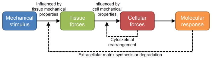
Mechanical forces direct cellular activities to induce tissue adaptation. Extrinsically and intrinsically generated mechanical forces load musculoskeletal tissues, with the characteristics of the resultant tissue forces being dependent on the ability of the tissue to resist those forces. Tissue forces are transmitted to the micromechanical environment of resident cells, with cellular mechanical properties influencing the characteristics of the cellular forces. Cells can modify their micromechanical environment via cytoskeletal rearrangement, which feedbacks to alter cellular sensitivity to incoming forces. When cellular forces are sufficient, the cell initiates a molecular response, which ultimately alters synthesis and/or degradation of the extracellular matrix. The latter alters tissue mechanical properties, which feeds back to influence tissue forces. (Reprinted from Thompson et al, by permission of Oxford University Press and the American Physical Therapy Association.)

A variety of extracellular receptors activate an overlapping network of mechanosensitive pathways. (A) Musculoskeletal cells can sense incoming mechanical signals using a diverse group of transmembrane mechanosensitive proteins (‘mechanosensors’), including stretch-activated ion channels, cell-membrane spanning G protein-coupled receptors, growth-factor receptors and integrins. The mechanical stimulation of these proteins can lead to changes in their affinity to binding partners or ion conductivity. (B) Mechanical stimulation of the mechanosensors and alteration in their binding capacity or ion conductivity converts the mechanical signal into a biochemical signal (‘biochemical coupling’) triggering intracellular signalling cascades. Many of the pathways overlap sharing signalling molecules. The convergence of the pathways results in the activation of select transcription factors, including nuclear factor of activated T cells, nuclear factor-κβ, activator protein 1, GATA4 (a member of the transcription factor family characterised by the ability to bind the DNA sequence ‘GATA’) and signal transducer and activator of transcription factors. The transcription factors translocate to the nucleus and modulate the expression of a panel of mechanosensitive genes, including early growth response 1, lex1, Fos, Jun and cyclooxygenase-2. Ultimately, the net sum of gene-expression reprogramming determines the functional response of the cell to a mechanical stimulus. Akt/PKB, protein kinase B; AP1, activator protein 1; CaMK, calcium/calmodulin-dependent kinase; Cox2, cyclooxygenase-2; DAG, diacyl-glycerol; Egr1, early growth response 1; ERK, extracellular signal-regulated kinase; FAK, focal adhesion kinase; IP3, inositol triphosphate; JNKs, c-Jun N-terminal kinases; MEK, mitogen-activated protein kinase; MEKK, mitogen-activated protein kinase kinase; MLCK, myosin light-chain kinase; NFAT, nuclear factor of activated T cells; NF-κβ, nuclear factor-κβ; NO, nitric oxide; NOS, nitric oxide synthase; PAK, p21-activated kinase; PI3K, phosphoinositide 3-kinase; PKC, protein kinase C; PLC, phospholipase C; Raf, rapidly accelerated fibrosarcoma kinase; Ras, rat sarcoma small GTPase; STATs, signal transducer and activator of transcription factors. (Reprinted from Thompson et al, by permission of Oxford University Press and the American Physical Therapy Association.)
Comment on
- Br J Sports Med.
Similar articles
-
Mechanotherapy: revisiting physical therapy and recruiting mechanobiology for a new era in medicine.Trends Mol Med. 2013 Sep;19(9):555-64. doi: 10.1016/j.molmed.2013.05.005. Epub 2013 Jun 18. Trends Mol Med. 2013. PMID: 23790684 Review.
-
Understanding Mechanobiology: Physical Therapists as a Force in Mechanotherapy and Musculoskeletal Regenerative Rehabilitation.Phys Ther. 2016 Apr;96(4):560-9. doi: 10.2522/ptj.20150224. Epub 2015 Dec 4. Phys Ther. 2016. PMID: 26637643 Free PMC article.
-
Invited Review for 20th Anniversary Special Issue of PLRev "AI for Mechanomedicine".Phys Life Rev. 2024 Dec;51:328-342. doi: 10.1016/j.plrev.2024.10.010. Epub 2024 Oct 24. Phys Life Rev. 2024. PMID: 39489078 Review.
-
Synthetic mechanobiology: engineering cellular force generation and signaling.Curr Opin Biotechnol. 2016 Aug;40:82-89. doi: 10.1016/j.copbio.2016.03.004. Epub 2016 Mar 26. Curr Opin Biotechnol. 2016. PMID: 27023733 Free PMC article. Review.
-
The cellular mechanobiology of aging: from biology to mechanics.Ann N Y Acad Sci. 2021 May;1491(1):3-24. doi: 10.1111/nyas.14529. Epub 2020 Nov 24. Ann N Y Acad Sci. 2021. PMID: 33231326 Free PMC article. Review.
Cited by
-
Mechanical stimulation of human dermal fibroblasts regulates pro-inflammatory cytokines: potential insight into soft tissue manual therapies.BMC Res Notes. 2020 Aug 27;13(1):400. doi: 10.1186/s13104-020-05249-1. BMC Res Notes. 2020. PMID: 32854782 Free PMC article.
-
Optimization of Tuina rolling manipulation parameters to promote blood circulation using a circulatory orthogonal experiment.J Phys Ther Sci. 2024 May;36(5):294-302. doi: 10.1589/jpts.36.294. Epub 2024 May 1. J Phys Ther Sci. 2024. PMID: 38694003 Free PMC article.
-
Kettlebell training in clinical practice: a scoping review.BMC Sports Sci Med Rehabil. 2019 Sep 3;11:19. doi: 10.1186/s13102-019-0130-z. eCollection 2019. BMC Sports Sci Med Rehabil. 2019. PMID: 31497302 Free PMC article.
-
Mechanotransduction of stem cells for tendon repair.World J Stem Cells. 2020 Sep 26;12(9):952-965. doi: 10.4252/wjsc.v12.i9.952. World J Stem Cells. 2020. PMID: 33033557 Free PMC article. Review.
-
Loss of the auxiliary α2δ1 voltage-sensitive calcium channel subunit impairs bone formation and anabolic responses to mechanical loading.JBMR Plus. 2024 Jan 10;8(2):ziad008. doi: 10.1093/jbmrpl/ziad008. eCollection 2024 Feb. JBMR Plus. 2024. PMID: 38505532 Free PMC article.
References
Publication types
MeSH terms
Grants and funding
LinkOut - more resources
Full Text Sources
Other Literature Sources