. 2017 Jun 23;7(1):4172.
doi: 10.1038/s41598-017-04075-z.

Breast Cancer Multi-classification from Histopathological Images with Structured Deep Learning Model

Affiliations

Breast Cancer Multi-classification from Histopathological Images with Structured Deep Learning Model

Zhongyi Han et al. Sci Rep. .

Abstract

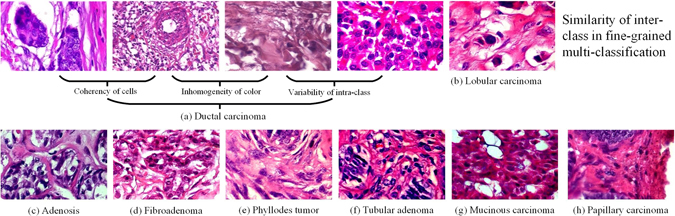
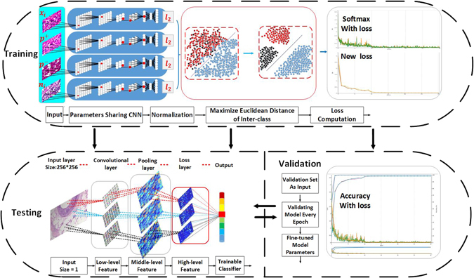
Automated breast cancer multi-classification from histopathological images plays a key role in computer-aided breast cancer diagnosis or prognosis. Breast cancer multi-classification is to identify subordinate classes of breast cancer (Ductal carcinoma, Fibroadenoma, Lobular carcinoma, etc.). However, breast cancer multi-classification from histopathological images faces two main challenges from: (1) the great difficulties in breast cancer multi-classification methods contrasting with the classification of binary classes (benign and malignant), and (2) the subtle differences in multiple classes due to the broad variability of high-resolution image appearances, high coherency of cancerous cells, and extensive inhomogeneity of color distribution. Therefore, automated breast cancer multi-classification from histopathological images is of great clinical significance yet has never been explored. Existing works in literature only focus on the binary classification but do not support further breast cancer quantitative assessment. In this study, we propose a breast cancer multi-classification method using a newly proposed deep learning model. The structured deep learning model has achieved remarkable performance (average 93.2% accuracy) on a large-scale dataset, which demonstrates the strength of our method in providing an efficient tool for breast cancer multi-classification in clinical settings.


Conflict of interest statement

The authors declare that they have no competing interests.

Figures

Figure 1
Figure 1
Eight classes of breast cancer histopathological images from BreaKHis dataset. There are great challenging histopathological images due to the broad variability of high-resolution image appearances, high coherency of cancerous cells, and extensive inhomogeneity of color distribution. These histopathological images were all acquired at a magnification factor of 400.
Figure 2
Figure 2
Overview of the integrated workflow. The overall approach of our method is composed of three stages: training, validation, and testing. The goal of the training stage is to learn the sufficient feature representation and optimize the distance of different classes’ feature space. The validation stage aims to fine-tune parameters and select models of each epoch. The testing stage is designed to evaluate the performance of the CSDCNN.
Figure 3
Figure 3
Multi-classification performance with recognition rates of the CSDCNN among patient level (PL) and image level (IL). Our method takes advantage of newly network structures, fast convergence rates, and strong generalization capabilities. These can be demonstrated by the validation set and testing set having almost the same accuracy.
Figure 4
Figure 4
The comparison between CSDCNN training from transfer learning (TL) and from scratch (FC) among patient level (PL) and image level (IL).
Figure 5
Figure 5
High-resolution breast cancer histopathological images labeled by pathologists. In practice, the region of the cancerization is only a fraction of histopathological images. The area separated by the yellow boxes represents the region of interest labeled by pathologists, which is always solely the region of cancerization.

References

    1. Collins FS, Varmus H. A new initiative on precision medicine. The New England journal of medicine. 2015;372 9:793–5. doi: 10.1056/NEJMp1500523. - DOI - PMC - PubMed
    1. Reardon S. Precision-medicine plan raises hopes. Nature. 2015;517 7536:540. doi: 10.1038/nature.2015.16774. - DOI - PubMed
    1. Stewart, B. W. & Wild, C. World cancer report 2014. international agency for research on cancer. World Health Organization505 (2014).
    1. Zheng, Y. et al. De-enhancing the dynamic contrast-enhanced breast mri for robust registration. In International Conference on Medical Image Computing and Computer-Assisted Intervention, 933–941 (Springer, 2007). - PMC - PubMed
    1. Cai Y, et al. Multi-modal vertebrae recognition using transformed deep convolution network. Computerized Medical Imaging and Graphics. 2016;51:11–19. doi: 10.1016/j.compmedimag.2016.02.002. - DOI - PubMed

Publication types