. 2017 Jun 23;7(1):4202.
doi: 10.1038/s41598-017-04097-7.

A microRNA signature of response to erlotinib is descriptive of TGFβ behaviour in NSCLC

Affiliations

A microRNA signature of response to erlotinib is descriptive of TGFβ behaviour in NSCLC

Madeline Krentz Gober et al. Sci Rep. .

Abstract

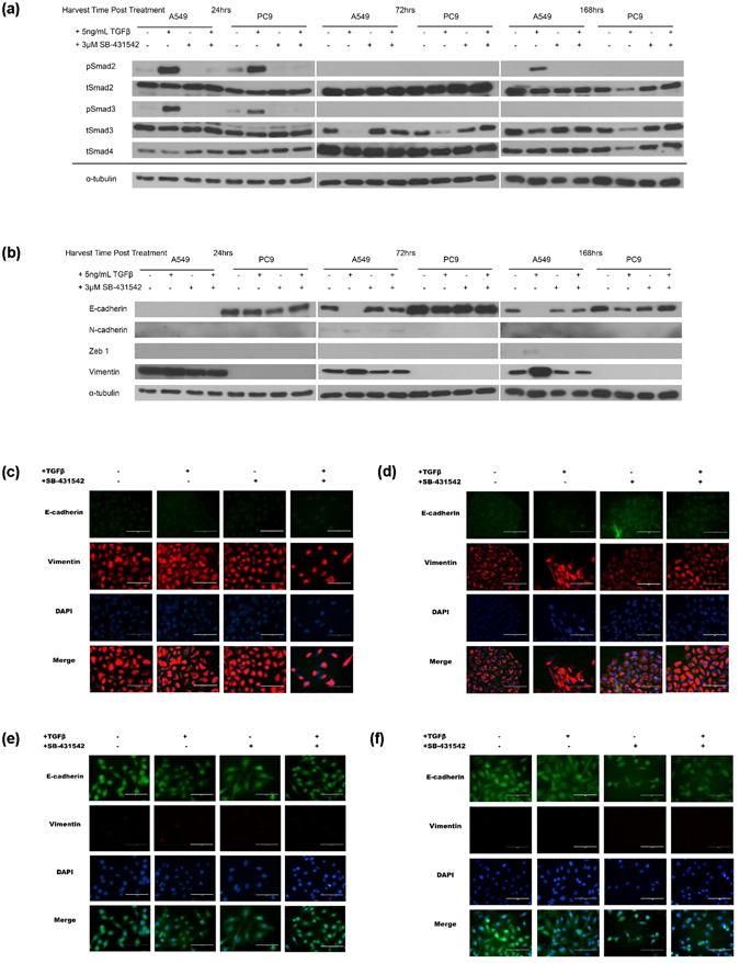
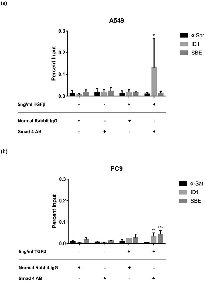
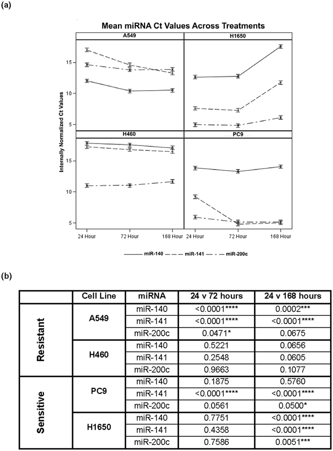
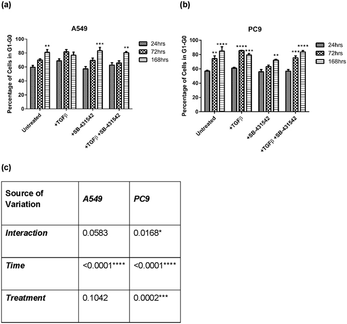
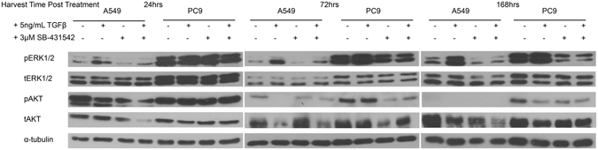
Our previous work identified a 13-gene miRNA signature predictive of response to the epidermal growth factor receptor (EGFR) inhibitor, erlotinib, in Non-Small Cell Lung Cancer cell lines. Bioinformatic analysis of the signature showed a functional convergence on TGFβ canonical signalling. We hypothesized that TGFβ signalling controls expression of the miRNA genes comprising an erlotinib response signature in NSCLC. Western analysis revealed that TGFβ signalling via Smad2/3/4 occurred differently between erlotinib-resistant A549 and erlotinib- sensitive PC9 cells. We showed that TGFβ induced an interaction between Smad4 and putative Smad Binding Elements in PC9. However, qRT-PCR analysis showed that endogenous miR-140/141/200c expression changes resulted from time in treatments, not the treatments themselves. Moreover, flow cytometry indicated that cells exited the cell cycle in the same manner. Taken together these data indicated that the miRNA comprising the signature are likely regulated by the cell cycle rather than by TGFβ. Importantly, this work revealed that TGFβ did not induce EMT in PC9 cells, but rather TGFβ-inhibition induced an EMT-intermediate. These data also show that growth/proliferation signals by constitutively-activated EGFR may rely on TGFβ and a possible relationship between TGFβ and EGFR signalling may prevent EMT progression in this context rather than promote it.


Conflict of interest statement

The authors declare that they have no competing interests.

Figures

Figure 1
Figure 1
Signature microRNA genes contain SBE elements. Promoter analysis was conducted using the ChipMAPPER algorithm, . microRNA genes -140, -141, and -200 contain putative SBE elements as represented by the triangle with conservative E-values less than or equal to 25 and a score greater than 3.0.
Figure 2
Figure 2
Total Smad expression, Smad activation and EMT program marker expression varies with TGFβ or inhibitor treatment. A549 and PC9 cells were plated, treated and harvested as described. Proteins were visualized by western blotting. α-tubulin levels are representative controls. (a) Profiling of Smad family member expression and activation across time demonstrates changes in TGFβ canonical signalling. (b) EMT protein markers demonstrate program initiation and progression among treatment conditions. (c) A549 cells treated for 24 hours for E-cadherin and Vimentin expression by immunofluorescence (d) A549 cells treated for 7 days for E-cadherin and Vimentin expression by immunofluorescence (e) PC9 cells treated for 24 hours for E-cadherin and Vimentin expression by immunofluorescence (f) PC9 cells treated for 7 days for E-cadherin and Vimentin expression by immunofluorescence.
Figure 3
Figure 3
TGFβ induces Smad4 binding to SBEs in the promoter of mir-200/141 in PC9 cells. Chromatin immunoprecipitation was performed to identify whether a physical interaction between Smad4 and a predicted SBE locus in the shared promoter of mir-200c/-141 resulted from TGFβ treatment. Normal rabbit IgG served as the antibody negative control and α-Satellite primers as the negative PCR control. ID1 locus immunoprecipitation was the positive control for Smad4 binding. (a) In A549, positive Smad4-ID1 association is observed with TGFβ treatment, but an Smad4-SBE interaction is not. (b) In PC9, both Smad4-ID1 and Smad4-SBE interaction is observed. Significance was calculated using an unpaired t-test comparing TGFβ-treated cells and -untreated samples with the same primer set.
Figure 4
Figure 4
Time of TGFβ treatment reflects changes in endogenous miRNA gene expression. Changes in endogenous gene expression were analysed using a five-way ANOVA considering the variables: TGFβ treatment, SB-431542 treatment, time point, expression as internally normalized Ct values, and cell line, along with all interaction terms. (a) Data presented here is aggregated by averaging over treatments in order to capture overarching trends in miRNA and cell line patterns at multiple time points. Fine-scale trends were broken down by individual treatments as presented in Supplemental Figure 4. (b) Comparison of the significance of endogenous expression changes between time points samples and by individual miRNA genes in each cell line.
Figure 5
Figure 5
A549 and PC9 cells exit the cell cycle regardless of treatment with TGFβ or SB-431542. The graph reflects the percentage of (a) A549 or (b) PC9 cell populations in G0-G1 phase of the cell cycle at 24, 72, and 168 hours following treatment. Significance was determined using an unpaired t-test comparing the 72 and 168 hour time points individually to the 24 hour time point of the same treatment. (c) A two-way ANOVA was utilized to determine the significance of treatment and/or time point reflective of the percentage of cells in the G0-G1 phase of the cell cycle.
Figure 6
Figure 6
TGFβ modulation differentially impacts ERK and AKT activation between A549 and PC9. A549 and PC9 cells were plated, treated, and harvested as described in the methods. α-tubulin levels are representative of an individual lysate pool. Lysates profiled here are the same as in Fig. 2. ERK-MAPK and PI3K-AKT signaling are non-canonical signaling effectors of the TGFβ signaling pathway.

References

    1. Kwak EL, et al. Anaplastic lymphoma kinase inhibition in non-small-cell lung cancer. N Engl J Med. 2010;363:1693–1703. doi: 10.1056/NEJMoa1006448. - DOI - PMC - PubMed
    1. Bergethon K, et al. ROS1 rearrangements define a unique molecular class of lung cancers. Journal of clinical oncology: official journal of the American Society of Clinical Oncology. 2012;30:863–870. doi: 10.1200/JCO.2011.35.6345. - DOI - PMC - PubMed
    1. The Cancer Genome Atlas Research, N. Comprehensive molecular profiling of lung adenocarcinoma. Nature 511, 543–550, doi:10.1038/nature13385http://www.nature.com/nature/journal/v511/n7511/abs/nature13385.html#sup... (2014). - PMC - PubMed
    1. Kim ES, et al. The BATTLE trial: personalizing therapy for lung cancer. Cancer discovery. 2011;1:44–53. doi: 10.1158/2159-8274.CD-10-0010. - DOI - PMC - PubMed
    1. Pao W, Miller VA. Epidermal growth factor receptor mutations, small-molecule kinase inhibitors, and non-small-cell lung cancer: current knowledge and future directions. Journal of clinical oncology: official journal of the American Society of Clinical Oncology. 2005;23:2556–2568. doi: 10.1200/JCO.2005.07.799. - DOI - PubMed

MeSH terms