. 2017 Aug;18(4):601-617.
doi: 10.1007/s10162-017-0628-6. Epub 2017 Jun 23.

Rhesus Cochlear and Vestibular Functions Are Preserved After Inner Ear Injection of Saline Volume Sufficient for Gene Therapy Delivery

Affiliations

Rhesus Cochlear and Vestibular Functions Are Preserved After Inner Ear Injection of Saline Volume Sufficient for Gene Therapy Delivery

Chenkai Dai et al. J Assoc Res Otolaryngol. 2017 Aug.

Abstract

Sensorineural losses of hearing and vestibular sensation due to hair cell dysfunction are among the most common disabilities. Recent preclinical research demonstrates that treatment of the inner ear with a variety of compounds, including gene therapy agents, may elicit regeneration and/or repair of hair cells in animals exposed to ototoxic medications or other insults to the inner ear. Delivery of gene therapy may also offer a means for treatment of hereditary hearing loss. However, injection of a fluid volume sufficient to deliver an adequate dose of a pharmacologic agent could, in theory, cause inner ear trauma that compromises functional outcome. The primary goal of the present study was to assess that risk in rhesus monkeys, which closely approximates humans with regard to middle and inner ear anatomy. Secondary goals were to identify the best delivery route into the primate ear from among two common surgical approaches (i.e., via an oval window stapedotomy and via the round window) and to determine the relative volumes of rhesus, rodent, and human labyrinths for extrapolation of results to other species. We measured hearing and vestibular functions before and 2, 4, and 8 weeks after unilateral injection of phosphate-buffered saline vehicle (PBSV) into the perilymphatic space of normal rhesus monkeys at volumes sufficient to deliver an atoh1 gene therapy vector. To isolate effects of injection, PBSV without vector was used. Assays included behavioral observation, auditory brainstem responses, distortion product otoacoustic emissions, and scleral coil measurement of vestibulo-ocular reflexes during whole-body rotation in darkness. Three groups (N = 3 each) were studied. Group A received a 10 μL transmastoid/trans-stapes injection via a laser stapedotomy. Group B received a 10 μL transmastoid/trans-round window injection. Group C received a 30 μL transmastoid/trans-round window injection. We also measured inner ear fluid space volume via 3D reconstruction of computed tomography (CT) images of adult C57BL6 mouse, rat, rhesus macaque, and human temporal bones (N = 3 each). Injection was well tolerated by all animals, with eight of nine exhibiting no signs of disequilibrium and one animal exhibiting transient disequilibrium that resolved spontaneously by 24 h after surgery. Physiologic results at the final, 8-week post-injection measurement showed that injection was well tolerated. Compared to its pretreatment values, no treated ear's ABR threshold had worsened by more than 5 dB at any stimulus frequency; distortion product otoacoustic emissions remained detectable above the noise floor for every treated ear (mean, SD and maximum deviation from baseline: -1.3, 9.0, and -18 dB, respectively); and no animal exhibited a reduction of more than 3 % in vestibulo-ocular reflex gain during high-acceleration, whole-body, passive yaw rotations in darkness toward the treated side. All control ears and all operated ears with definite histologic evidence of injection through the intended site showed similar findings, with intact hair cells in all five inner ear sensory epithelia and intact auditory/vestibular neurons. The relative volumes of mouse, rat, rhesus, and human inner ears as measured by CT were (mean ± SD) 2.5 ± 0.1, 5.5 ± 0.4, 59.4 ± 4.7 and 191.1 ± 4.7 μL. These results indicate that injection of PBSV at volumes sufficient for gene therapy delivery can be accomplished without destruction of inner ear structures required for hearing and vestibular sensation.

Keywords: auditory brainstem response; gene therapy; inner ear; otoacoustic emission; safety; vestibular ocular reflex; volume.


Figures

Fig. 1
Fig. 1
Auditory brainstem response (ABR) thresholds in response to clicks and pure tones at 1, 2, 4 and 8 kHz of three monkeys/group in three treatment groups. Top row: group A (right ear transmastoid/trans-oval window/stapes [TM/OW/Stapes] injection of 10 μL phosphate-buffered saline vehicle [PBSV] via a stapedotomy). Middle row: group B (right ear transmastoid/trans-round window [TM/RW] injection of 10 μL PBSV). Bottom row: group C (right ear TM/RW injection of 30 μL PBSV). N and black triangles/error bars: mean ± SD thresholds measured in 18 normal rhesus ears before any manipulation. L left/untreated; R right/treated; 0 before injection; 2, 4, 8 number of weeks after right inner ear PBSV injection. Threshold units are dB HL as reported by a clinical ABR system using the human A-weighted scale. Monkey 24X=formula image, 73X=formula image, 66X=formula image, 32X=formula image, 43X=formula image, 12X=formula image, 36X=formula image, 37X=formula image, 49X=formula image.
Fig. 2
Fig. 2
DP-NF (i.e., DPOAE amplitudes relative to noise floor amplitude, in dB) atf2 = 1, 2, 4, and 8 kHz for the same treatment conditions, animals, ears, and time points shown in Fig. 1. Monkey 24X=formula image, 73X=formula image, 66X=formula image, 32X=formula image, 43X=formula image, 12X=formula image, 36X=formula image, 37X=formula image, 49X=formula image
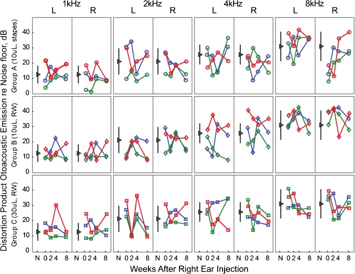
. A DP-NF value >0 indicates presence of functional outer hair cells in cochlea.
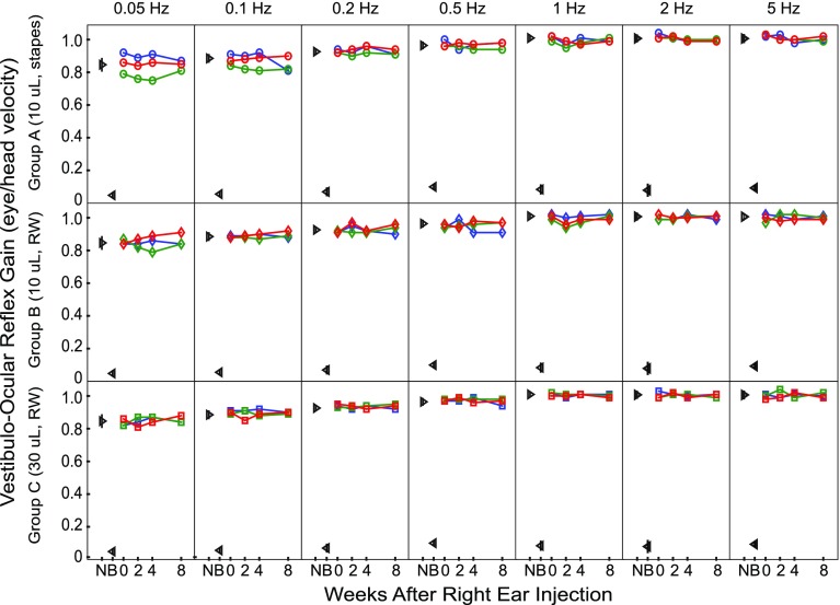
Fig. 3
Fig. 3
VOR Gain [eye velocity/head velocity] during eye movement responses to passive sinusoidal whole-body rotation in darkness at frequencies of 0.05 to 5 Hz. N normal, mean ± SD VOR gain measured in all nine monkeys before inner ear injection; B VOR gain data from (Dai et al. 2012) for four rhesus monkeys with bilateral vestibular deficiency after bilateral ototoxic injury; 0 before injection; 2, 4, 8 number of weeks after inner ear PBSV injection. Monkey 24X=formula image, 73X=formula image, 66X=formula image, 32X=formula image, 43X=formula image, 12X=formula image, 36X=formula image, 37X=formula image, 49X=formula image. SD <0.03 when SD bars are not visible for N and B data.
Fig. 4
Fig. 4
Mean ± SD VOR “acceleration gain” GA [eye velocity/head velocity, averaged over the constant-acceleration segment of the head movement] for whole-body, passive 1000 °/s2 impulse head rotations in darkness before (0) and at 2, 4, and 8 weeks after right inner ear PBSV injection. N Normal, B bilateral vestibular deficiency due to intratympanic administration of gentamicin (Dai et al. 2011b). Left panels show data for leftward head turns, predominantly reflecting left (non-injected ear) horizontal semicircular canal function. Right panels show rightward rotation data, predominantly reflecting right (treated ear) horizontal semicircular canal function. Monkey 24X=formula image, 73X=formula image, 66X=formula image, 32X=formula image, 43X=formula image, 12X=formula image, 36X=formula image, 37X=formula image, 49X=formula image. SD <0.03 when SD bars are not visible beyond markers for N and B data. RW injected via round window, OW injected via oval window (stapes).
Fig. 5
Fig. 5
Representative hematoxylin and eosin stained light microscopy images of treated right ears. a In an animal that underwent oval window injection (group A monkey 24X right ear), the mastoid exhibits fibrous tissue and minimal inflammation after surgical mastoidectomy, with preservation of the horizontal SCC (H), facial nerve (FN), incus (I), and tympanic membrane (TM). b In the same ear, a part of the stapes footplate has been replaced by a thin fibrous scar band (arrow), indicating the site of catheter penetration and injection. Blood products are visible with occasional mononuclear cells. c In an animal that underwent round window injection (group B monkey 12X right ear), the round window (arrow head) appears to be intact without evidence of fibrosis or fenestration. Blood products are visible in the round window niche.
Fig. 6
Fig. 6
Representative hematoxylin and eosin stained light microscopy images of the cochleae in a normal ear (a, e monkey 37X left ear) and the treated right ears from animals in group A (b, f monkey 24X), group B (c g monkey 12X), and group C (d, h monkey 36X). Mid-modiolar sections (ad, 2.5X) show intact basal, mid, and apical turns in normal ear and all treatment groups. High magnification images of the organ of Corti for the same specimens (eh 20X) show variable disruption of Reissner’s membrane (RM) but the presence of intact inner (arrow head) and outer hair cells (arrow) and cochlear nerve fibers. ST scala tympani, SM scala media, SV scala vestibuli.
Fig. 7
Fig. 7
Representative light microscopy images of cristae (a, d, g, j), utricles (b, e, h, k), and saccules (c, f, i, l) in three study ears, one from each group. ac Monkey 73X left ear, normal. df Monkey 73X right ear, group A. gi Monkey 32X right ear, group B. jl Monkey 36X right ear, group C. Although histologic artifacts and differences in the plane of section are apparent, all specimens exhibit intact hair cells in the neuroepithelia, intact neurons, and absence of intralumenal fibrosis or signs of infection.
Fig. 8
Fig. 8
Inner ear fluid space volume surface reconstructions from micro-computed tomography scans of a C57BL/6 J mouse, b Wistar rat, c rhesus monkey, and d human.

References

    1. Akil O, Rouse SL, Chan DK, Lustig LR (2015) Surgical method for virally mediated gene delivery to the mouse inner ear through the round window membrane. J Vis Exp 16(97). doi:10.3791/52187 - PMC - PubMed
    1. Borenstein JT. Intracochlear drug delivery systems. Expert Opin Drug Deliv. 2011;8(9):1161–1174. doi: 10.1517/17425247.2011.588207. - DOI - PMC - PubMed
    1. Brown RF, Hullar TE, Cadieux JH, Chole RA. Residual hearing preservation after pediatric cochlear implantation. Otol Neurotol. 2010;31(8):1221–1216. doi: 10.1097/MAO.0b013e3181f0c649. - DOI - PMC - PubMed
    1. Bruce IA, Bates JE, Melling C, Mawman D, Green KM. Hearing preservation via a cochleostomy approach and deep insertion of a standard length cochlear implant electrode. Otol Neurotol. 2011;32(9):1444–14447. doi: 10.1097/MAO.0b013e3182355824. - DOI - PubMed
    1. Buckingham RA, Valvassori GE. Inner ear fluid volumes and the resolving power of magnetic resonance imaging: can it differentiate endolymphatic structures? Ann Otol Rhinol Laryngol. 2001;110(2):113–117. doi: 10.1177/000348940111000204. - DOI - PubMed

Publication types