. 2018 Apr;34(2):261-269.
doi: 10.1007/s12264-017-0148-8. Epub 2017 Jun 23.

Tau-Induced Ca2+/Calmodulin-Dependent Protein Kinase-IV Activation Aggravates Nuclear Tau Hyperphosphorylation

Affiliations

Tau-Induced Ca2+/Calmodulin-Dependent Protein Kinase-IV Activation Aggravates Nuclear Tau Hyperphosphorylation

Yu-Ping Wei et al. Neurosci Bull. 2018 Apr.

Abstract

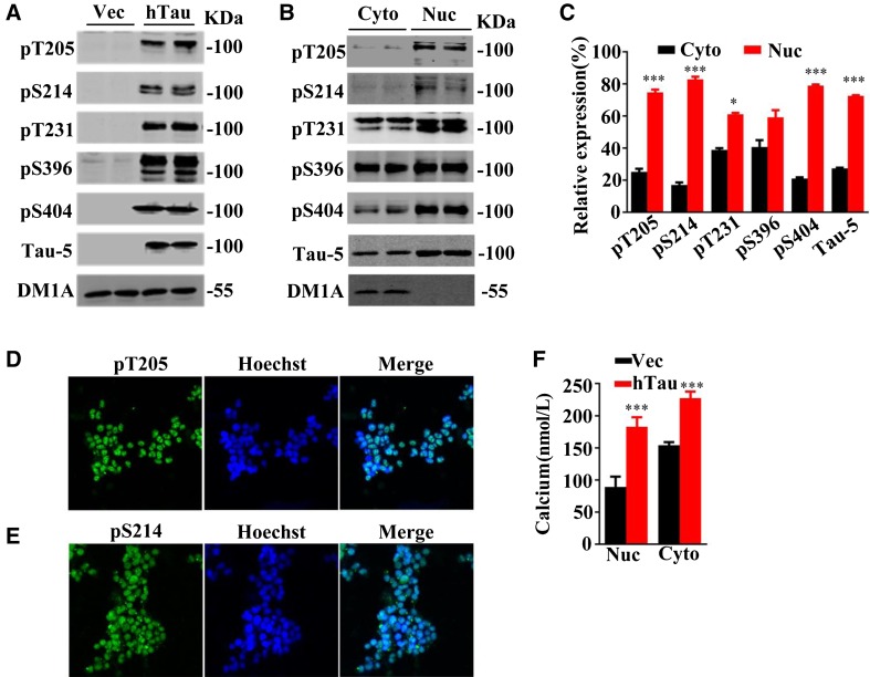
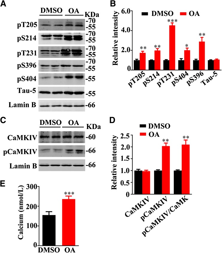
Hyperphosphorylated tau is the major protein component of neurofibrillary tangles in the brains of patients with Alzheimer's disease (AD). However, the mechanism underlying tau hyperphosphorylation is not fully understood. Here, we demonstrated that exogenously expressed wild-type human tau40 was detectable in the phosphorylated form at multiple AD-associated sites in cytoplasmic and nuclear fractions from HEK293 cells. Among these sites, tau phosphorylated at Thr205 and Ser214 was almost exclusively found in the nuclear fraction at the conditions used in the present study. With the intracellular tau accumulation, the Ca2+ concentration was significantly increased in both cytoplasmic and nuclear fractions. Further studies using site-specific mutagenesis and pharmacological treatment demonstrated that phosphorylation of tau at Thr205 increased nuclear Ca2+ concentration with a simultaneous increase in the phosphorylation of Ca2+/calmodulin-dependent protein kinase IV (CaMKIV) at Ser196. On the other hand, phosphorylation of tau at Ser214 did not significantly change the nuclear Ca2+/CaMKIV signaling. Finally, expressing calmodulin-binding protein-4 that disrupts formation of the Ca2+/calmodulin complex abolished the okadaic acid-induced tau hyperphosphorylation in the nuclear fraction. We conclude that the intracellular accumulation of phosphorylated tau, as detected in the brains of AD patients, can trigger nuclear Ca2+/CaMKIV signaling, which in turn aggravates tau hyperphosphorylation. Our findings provide new insights for tauopathies: hyperphosphorylation of intracellular tau and an increased Ca2+ concentration may induce a self-perpetuating harmful loop to promote neurodegeneration.

Keywords: Alzheimer’s disease; CaMKIV; Nuclear calcium signal; Phosphorylation; Tau.


Figures

Fig. 1
Fig. 1
Expression of phosphorylated tau increases Ca2+ in cytoplasm and nucleus. A Western blots of extracts from HEK293 cells transiently transfected with DsRed2-N1-human tau40 plasmid for 24 h, showing phosphorylation at Thr205, Ser214, Thr231, Ser396, and Ser404 and total tau (probed by Tau-5). B, C Western blots and levels of tau proteins in the cytoplasmic (Cyto) and nuclear (Nuc) fractions. D, E Representative images showing co-localization of Thr205- (pT205) and Ser214- (pS214) phosphorylated tau with a nuclear marker (Hoechst) after transfection in HEK293 cells. F Basal [Ca2+] increased in the nucleus and cytoplasm of HEK293 cells transfected with human wild-type tau40 compared with vehicle (Vec) for 24 h (>100 cells). Data are expressed as mean ± SEM; *P < 0.05, ***P < 0.001 vs Vec or Cyto.
Fig. 2
Fig. 2
Phosphorylation of tau at Thr205 activates the nuclear Ca2+-CaMKIV pathway. A, B HEK293 cells were transfected with DsRed2-N1-phospho-mimic (T205E) or non-phospho-mimic (T205A) mutant tau plasmids for 24 h and then the nuclear fraction was prepared for Western blotting. An increased phosphorylation level of CaMKIV was detected in the nuclear fraction of T205E-transfected cells compared with T205A. C An increased basal [Ca2+] was detected in the nuclear fraction of T205E-transfected cells compared with T205A (>100 cells). Data are expressed as mean ± SEM; *P < 0.05, **P < 0.01, ***P < 0.001 vs T205A.
Fig. 3
Fig. 3
Phosphorylation of tau at Ser214 does not affect nuclear Ca2+ and CaMKIV activity. A, B HEK293 cells were transfected with DsRed2-N1-phospho-mimic (S214E) or non-phospho-mimic (S214A) mutant tau plasmids for 24 h and then the nuclear fraction was prepared for Western blotting. Compared with S214A, expression of S214E did not significantly change the levels of total CaMKIV and pCaMKIV in the nuclear fraction. C Compared with S214A, expression of S214E did not change the level of nuclear Ca2+. Data are expressed as mean ± SEM.
Fig. 4
Fig. 4
OA induces tau hyperphosphorylation with nuclear Ca2+/CaMKIV upregulation. A, B HEK293 cells with stable expression of human wild-type tau (293Tau) were treated with OA (10 nmol/L) for 12 h, and the increased phosphorylation level of tau at Thr204, Ser214, Thr231, Ser396, and Ser404 was detected in the nuclear fraction by Western blotting (Tau5 probes total tau proteins). C, D OA treatment further increased pCaMKIV in the nuclear fraction. E OA treatment further increased nuclear [Ca2+]. Data are expressed as mean ± SEM; *P < 0.05, **P < 0.01, ***P < 0.001 vs OA.
Fig. 5
Fig. 5
Inhibition of CaMKIV abolishes OA-induced tau hyperphosphorylation in the nuclear fraction. A, B HEK293 cells with stable expression of human wild-type tau (293Tau) were transfected with CaMBP4 plasmid or vector for 24 h, and a decreased level of pCaMKIV in the nuclear fraction of the CaMBP4-trasfected cells was detected by Western blotting (**P < 0.01 vs Vec). C, D HEK293 cells with stable expression of human wild-type tau were transfected with CaMBP4 or the vector for 24 h, and then treated with OA or DMSO for 12 h. CaMBP4 expression reduced the OA-induced tau hyperphosphorylation at Thr205, Ser214, Thr213, Ser396, and Ser404 in the nuclear fraction. Tau-5 probes total tau proteins. Data are expressed as mean ± SEM; *P < 0.05, **P < 0.01, ***P < 0.001 vs Vec + DMSO; # P < 0.05, ## P < 0.01 vs Vec + OA.

References

    1. Grundke-Iqbal I, Iqbal K, Quinlan M, Tung YC, Zaidi MS, Wisniewski HM. Microtubule-associated protein tau. A component of Alzheimer paired helical filaments. J Biol Chem. 1986;261:6084–6089. - PubMed
    1. Grundke-Iqbal I, Iqbal K, Tung YC, Quinlan M, Wisniewski HM, Binder LI. Abnormal phosphorylation of the microtubule-associated protein tau (tau) in Alzheimer cytoskeletal pathology. Proc Natl Acad Sci U S A. 1986;83:4913–4917. doi: 10.1073/pnas.83.13.4913. - DOI - PMC - PubMed
    1. Masters CL, Simms G, Weinman NA, Multhaup G, McDonald BL, Beyreuther K. Amyloid plaque core protein in Alzheimer disease and Down syndrome. Proc Natl Acad Sci U S A. 1985;82:4245–4249. doi: 10.1073/pnas.82.12.4245. - DOI - PMC - PubMed
    1. Wang JZ, Wang ZH, Tian Q. Tau hyperphosphorylation induces apoptotic escape and triggers neurodegeneration in Alzheimer’s disease. Neurosci Bull. 2014;30:359–366. doi: 10.1007/s12264-013-1415-y. - DOI - PMC - PubMed
    1. Pierrot N, Santos SF, Feyt C, Morel M, Brion JP, Octave JN. Calcium-mediated transient phosphorylation of tau and amyloid precursor protein followed by intraneuronal amyloid-beta accumulation. J Biol Chem. 2006;281:39907–39914. doi: 10.1074/jbc.M606015200. - DOI - PubMed

LinkOut - more resources