. 2017 Sep:140:115-127.
doi: 10.1016/j.biomaterials.2017.06.004. Epub 2017 Jun 6.

A highly adhesive and naturally derived sealant

Affiliations

A highly adhesive and naturally derived sealant

Alexander Assmann et al. Biomaterials. 2017 Sep.

Abstract

Conventional surgical techniques to seal and repair defects in highly stressed elastic tissues are insufficient. Therefore, this study aimed to engineer an inexpensive, highly adhesive, biocompatible, and biodegradable sealant based on a modified and naturally derived biopolymer, gelatin methacryloyl (GelMA). We tuned the degree of gelatin modification, prepolymer concentration, photoinitiator concentration, and crosslinking conditions to optimize the physical properties and adhesion of the photocrosslinked GelMA sealants. Following ASTM standard tests that target wound closure strength, shear resistance, and burst pressure, GelMA sealant was shown to exhibit adhesive properties that were superior to clinically used fibrin- and poly(ethylene glycol)-based glues. Chronic in vivo experiments in small as well as translational large animal models proved GelMA to effectively seal large lung leakages without the need for sutures or staples, presenting improved performance as compared to fibrin glue, poly(ethylene glycol) glue and sutures only. Furthermore, high biocompatibility of GelMA sealant was observed, as evidenced by a low inflammatory host response and fast in vivo degradation while allowing for adequate wound healing at the same time. Combining these results with the low costs, ease of synthesis and application of the material, GelMA sealant is envisioned to be commercialized not only as a sealant to stop air leakages, but also as a biocompatible and biodegradable hydrogel to support lung tissue regeneration.

Keywords: Gelatin methacryloyl (GelMA); Hydrogel; Lung lesion; Sealant; Wound repair.


Figures

Figure 1
Figure 1. In vitro sealing properties of GelMA sealant
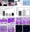
(a,b) Standard wound closure test using porcine skin as a biological substrate to test the in vitro adhesion strength of GelMA and different commercially available sealants. (c,d) Standard lap shear test to determine the shear strength of GelMA sealant in comparison to different commercially available sealants. The effect of GelMA prepolymer concentrations on the adhesion strength of hydrogels formed at 3 min UV exposure time is shown in a,c; the effect of the UV exposure time on the adhesion strength of a 25% GelMA sealant is shown in b,d. (*p<0.05, **p<0.01, ***p<0.001, ****p<0.0001).
Figure 2
Figure 2. In vitro burst pressure of GelMA sealant
Burst pressure values for commercially available sealants and GelMA sealants produced by (a,b) varying GelMA concentrations at 3 min UV exposure time and (c,d) changing UV exposure time for a 25% GelMA sealant. (**p<0.01; ***p<0.001; ****p<0.0001).
Figure 3
Figure 3. In vivo biocompatibility of GelMA sealant
Immunohistology analyses (a-c) 3, (d-f) 7, and (g-i) 28 days after subcutaneous implantation in rats showed initial implant-surrounding macrophage (CD68) invasion (arrows in b,e), which was no longer present at day 28 (h). At no point were there signs of lymphocyte (CD3) infiltration (c,f,i). (a,d,g, hematoxylin/eosin staining; asterisks, GelMA sealant; scale bars, 200 μm).
Figure 4
Figure 4. In vivo sealing capacity of GelMA sealant using a rat lung incision model
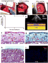
(a,b) GelMA sealant is applied on a lung leakage via a small lateral thoracotomy and UV-crosslinked until the incision was sealed. (c) Schematic of the setup used to measure the lung burst pressure after sealing: A syringe pump and a pressure sensor are connected to the trachea allowing for pressure monitoring during lung inflation in a closed system. (d) Burst pressure of GelMA-sealed, Evicel®-sealed, Progel™-sealed and sutured lungs immediately after material application demonstrating that the burst pressures of GelMA sealant-treated lungs were significantly higher than those in all other groups. (e) Burst pressure of GelMA-sealed lungs on day 0 and day 7 post surgery compared to healthy lung: 7 days after surgery, the burst pressure of GelMA-sealed lungs was further increased as compared to day 0 and reached the level of healthy lung tissue. (f,g) Hematoxylin/eosin and (h,k) masson trichrome stainings of GelMA-sealed lung tissue sections at days 7 and 28. Histologically, defect repair tissue was observed under the GelMA cover at day 7 after lung leakage sealing (asterisks in f), including stable collagenous tissue layers (asterisks in h). After 28 days in vivo, host cells had invaded the GelMA seal and matrix remodeling had occurred (hashs in g,k). Immunohistology of GelMA-sealed lung tissue sections at day 7 (m) and day 28 (n) revealed only sparse presence of CD3- and CD68-positive cells around the GelMA implants (asterisks in m, defect repair tissue; hashs in n, remodeled hydrogel cover). (scale bars, 100 μm and 50 μm in the high magnification pictures, respectively; **p<0.01; ***p<0.001).
Figure 5
Figure 5. In vivo sealing capacity of GelMA sealant using a porcine lung incision model
(a-c) A right lung lobe is exposed via a small lateral thoracotomy, a standardized defect is created (broken lines in a and b) and sealed by photocrosslinking of GelMA sealant (broken line in c). (d,e) Ultrasound studies on the sealed lung tissue at postoperative days 7 and 14: Freedom from pneumothorax was confirmed by sonography, as shown in a representative ultrasound image at day 14. (f-h) Representative histological sections of the GelMA-sealed site after 14 days revealed sufficient wound healing (asterisks in f) including a stable collagenous (blue) defect cover (asterisks in g), in comparison with non-injured parenchymatous lung tissue (h). For high magnification pictures, see Figure S9. (i) By immunohistological staining of the GelMA-sealed tissue, no lymphocyte infiltration (CD3) was observed, and only mild macrophage accumulation (Mac-2) occurred. (f, hematoxylin/eosin staining; g,h, masson trichrome staining; scale bars in f,g,h, 200 μm; scale bar in i, 100 μm).

References

    1. Itano H. The optimal technique for combined application of fibrin sealant and bioabsorbable felt against alveolar air leakage. Eur J Cardiothorac Surg. 2008;33:457–60. - PubMed
    1. Glickman M, Gheissari A, Money S, Martin J, Ballard JL, Surger CMV. A polymeric sealant inhibits anastomotic suture hole bleeding more rapidly than gelfoam/thrombin - Results of a randomized controlled trial. Arch Surg-Chicago. 2002;137:326–31. - PubMed
    1. Annabi N, Tamayol A, Shin SR, Ghaemmaghami AM, Peppas NA, Khademhosseini A. Surgical Materials: Current Challenges and Nano-enabled Solutions. Nano Today. 2014;9:574–89. - PMC - PubMed
    1. Annabi N, Yue K, Tamayol A, Khademhosseini A. Elastic sealants for surgical applications. European journal of pharmaceutics and biopharmaceutics : official journal of Arbeitsgemeinschaft fur Pharmazeutische Verfahrenstechnik eV. 2015;95:27–39. - PMC - PubMed
    1. Wolbank S, Pichler V, Ferguson JC, Meinl A, van Griensven M, Goppelt A, et al. Non-invasive in vivo tracking of fibrin degradation by fluorescence imaging. J Tissue Eng Regen Med. 2015;9:973–6. - PubMed

MeSH terms