. 2017 Jun 24;8(1):150.
doi: 10.1186/s13287-017-0601-7.

Modulation of oxidative phosphorylation and redox homeostasis in mitochondrial NDUFS4 deficiency via mesenchymal stem cells

Affiliations

Modulation of oxidative phosphorylation and redox homeostasis in mitochondrial NDUFS4 deficiency via mesenchymal stem cells

Marlen Melcher et al. Stem Cell Res Ther. .

Abstract

Background: Disorders of the oxidative phosphorylation (OXPHOS) system represent a large group among the inborn errors of metabolism. The most frequently observed biochemical defect is isolated deficiency of mitochondrial complex I (CI). No effective treatment strategies for CI deficiency are so far available. The purpose of this study was to investigate whether and how mesenchymal stem cells (MSCs) are able to modulate metabolic function in fibroblast cell models of CI deficiency.

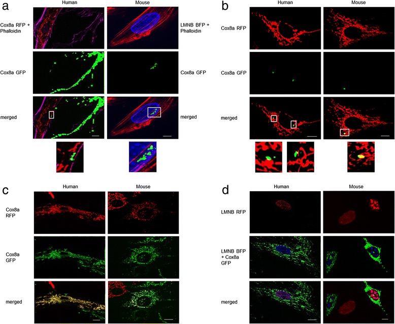
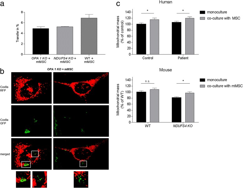
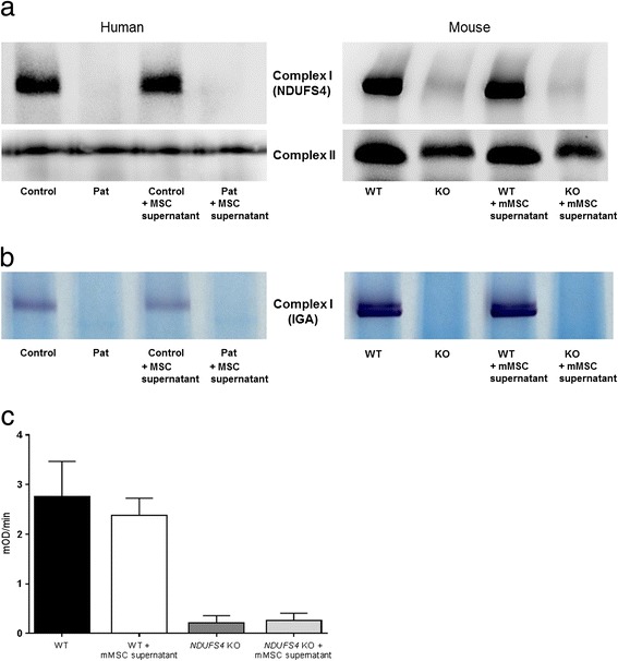
Methods: We used human and murine fibroblasts with a defect in the nuclear DNA encoded NDUFS4 subunit of CI. Fibroblasts were co-cultured with MSCs under different stress conditions and intercellular mitochondrial transfer was assessed by flow cytometry and fluorescence microscopy. Reactive oxygen species (ROS) levels were measured using MitoSOX-Red. Protein levels of CI were analysed by blue native polyacrylamide gel electrophoresis (BN-PAGE).

Results: Direct cellular interactions and mitochondrial transfer between MSCs and human as well as mouse fibroblast cell lines were demonstrated. Mitochondrial transfer was visible in 13.2% and 6% of fibroblasts (e.g. fibroblasts containing MSC mitochondria) for human and mouse cell lines, respectively. The transfer rate could be further stimulated via treatment of cells with TNF-α. MSCs effectively lowered cellular ROS production in NDUFS4-deficient fibroblast cell lines (either directly via co-culture or indirectly via incubation of cell lines with cell-free MSC supernatant). However, CI protein expression and activity were not rescued by MSC treatment.

Conclusion: This study demonstrates the interplay between MSCs and fibroblast cell models of isolated CI deficiency including transfer of mitochondria as well as modulation of cellular ROS levels. Further exploration of these cellular interactions might help to develop MSC-based treatment strategies for human CI deficiency.


Figures

Fig. 1
Fig. 1
a, b Mitochondrial transfer from MSCs to fibroblast cell lines. a Representative fluorescence images showing mitochondrial transfer and TNT formation. Mitochondria of stem cells are labelled with Cox8a GFP (green fluorescent protein), mitochondria of human fibroblasts are labelled with Cox8a RFP (red fluorescent protein) and nuclei of murine fibroblasts are labelled with LMNB BFP (blue fluorescent protein). Co-cultures were fixed and stained with phalloidin. b Confocal images of mitochondrial transfer derived from MSCs. Fibroblast mitochondria are labelled with Cox8a RFP and stem cell mitochondria are labelled with Cox8a GFP. c, d Representative fluorescence images of cell fusion between stem cell and fibroblast. c Mitochondria are labelled with Cox8a GFP (stem cell) and Cox8a RFP (fibroblast). Cells with a complete co-localization of red and green fluorescence indicate the rare event of nuclear fusion (yellow cells). d Stem cells have Cox8a GFP-labelled mitochondria and LMNB BFP-labelled nuclei. Fibroblasts have LMNB RFP-labelled nuclei. Cells with a co-localization of LMNB BFP and LMNB RFP have fused nuclei. Scale bars represent 10 μm. Images were contrast optimized (Colour figure online). Cox8a Cytochrome c oxidase subunit 8A, LMNB Lamin B1
Fig. 2
Fig. 2
Quantitative FACS measurement of mitochondrial transfer. a Flow cytometry analysis was performed after 48, 72 and 96 h of co-culture. Co-cultures between MSCs and fibroblasts and between fibroblasts and fibroblasts (e.g. healthy controls with NDUFS4-deficient cell lines) were measured. b Mitochondrial transfer during different culture conditions/drug treatment. mMSC mouse mesenchymal stem cell, MSC mesenchymal stem cell, Gal galactose medium, BSO buthioninesulfoximine, 2DG 2-deoxy-d-glucose, TNF-α tumour necrosis factor alpha, NDUFS4 NADH dehydrogenase ubiquinone Fe-S protein 4, WT wild type. Data shown as mean (% of “mito-positive” cells per measurement) ± SD. *p < 0.05, **p < 0.01, ***p < 0.001
Fig. 3
Fig. 3
Mitochondrial transfer between OPA1 KO fibroblasts and murine MSCs. a OPA1 KO fibroblasts were transfected with LMNB RFP (red fluorescent protein) to label the nuclei and mMSCs were transfected with Cox8a GFP (green fluorescent protein) to label the mitochondria. Flow cytometry was performed after 72 h co-culture. Data shown as mean (% of “mito-positive” cells per measurement) ± standard deviation. b Fluorescence images of mitochondrial transfer. Mitochondria of OPA1 KO fibroblasts were transfected with Cox8a RFP and stem cell mitochondria were transfected with Cox8a GFP. Scale bars represent 10 μm. Images were contrast optimized. c Mitochondrial mass was measured via live-cell microscopy in fibroblasts with and without visible mitochondrial transfer from MSCs. A significant increase was detected in fibroblasts that received MSC mitochondria during co-cultivation. *p < 0.05. n.s. not significant, mMSC mouse mesenchymal stem cell, MSC mesenchymal stem cell, NDUFS4 NADH dehydrogenase ubiquinone Fe-S protein 4, OPA1 optic atrophy 1, WT wild type (Colour figure online)
Fig. 4
Fig. 4
Mitochondrial ROS production was measured using MitoSOX Red (mitochondria-targeted superoxide indicator). a Fibroblast nuclei were labelled with LMNB BFP (blue fluorescent protein) and stem cell mitochondria were labelled with Cox8a GFP. Only fibroblasts with a blue fluorescent nucleus were measured. The measured fibroblasts were divided into two groups: fibroblasts with obviously transferred mitochondria derived from MSCs; and fibroblasts without transferred mitochondria. b MitoSOX measurement was performed after 72-h treatment with stem cell supernatant or control cell supernatant. Data shown as mean ± SEM. *p < 0.05, ***p < 0.001. c Representative fluorescence images showing a transferred MSC mitochondrion (green) in a human fibroblast (blue nuclei). Mitochondrial MitoSOX fluorescence is shown (red). Scale bar represents 10 μm. mMSC mouse mesenchymal stem cell, MSC mesenchymal stem cell, NDUFS4 NADH dehydrogenase ubiquinone Fe-S protein 4, ROS reactive oxygen species, WT wild type (Colour figure online)
Fig. 5
Fig. 5
a BN-PAGE with mitochondrial lysates from human and mouse fibroblasts cell lines shows no recovery of fully assembled complex I levels upon treatment with MSC supernatant. Complex II SDHA was used as loading control. b Complex I in-gel-activity assay demonstrates no improvement of CI activity upon treatment with MSC supernatant. c A complex I enzyme activity microplate assay kit was used to determine the complex I activity in mouse NDUFS4 KO and WT fibroblasts treated or untreated with supernatant from mMSCs. In accordance with BN-PAGE analysis, no significant increase in activity could be detected. Rate (mOD/min) = Absorbance 1 – Absorbance 2 / time (min). mMSC mouse mesenchymal stem cell, MSC mesenchymal stem cell, NDUFS4 NADH dehydrogenase ubiquinone Fe-S protein 4, WT wild type

References

    1. Kruse SE, Watt WC, Marcinek DJ, Kapur RP, Schenkman KA, Palmiter RD. Mice with mitochondrial complex I deficiency develop a fatal encephalomyopathy. Cell Metab. 2008;7(4):312–20. doi: 10.1016/j.cmet.2008.02.004. - DOI - PMC - PubMed
    1. Liu CS, Chang JC, Kuo SJ, Liu KH, Lin TT, Cheng WL, Chuang SF. Delivering healthy mitochondria for the therapy of mitochondrial diseases and beyond. Int J Biochem Cell Biol. 2014;53:141–6. doi: 10.1016/j.biocel.2014.05.009. - DOI - PubMed
    1. Spees JL, Olson SD, Whitney MJ, Prockop DJ. Mitochondrial transfer between cells can rescue aerobic respiration. Proc Natl Acad Sci U S A. 2006;103(5):1283–8. doi: 10.1073/pnas.0510511103. - DOI - PMC - PubMed
    1. Alam MT, Manjeri GR, Rodenburg RJ, Smeitink JA, Notebaart RA, Huynen M, Willems PH, Koopman WJ. Skeletal muscle mitochondria of NDUFS4-/- mice display normal maximal pyruvate oxidation and ATP production. Biochim Biophys Acta. 2015;1847:526–33. doi: 10.1016/j.bbabio.2015.02.006. - DOI - PubMed
    1. Distelmaier F, Valsecchi F, Liemburg-Apers DC, Lebiedzinska M, Rodenburg RJ, Heil S, Keijer J, Fransen J, Imamura H, Danhauser K, et al. Mitochondrial dysfunction in primary human fibroblasts triggers an adaptive cell survival program that requires AMPK-alpha. Biochim Biophys Acta. 2015;1852(3):529–40. doi: 10.1016/j.bbadis.2014.12.012. - DOI - PubMed