Review
. 2018 Jan 20;28(3):251-272.
doi: 10.1089/ars.2017.7216. Epub 2017 Jul 28.

NAD(H) and NADP(H) Redox Couples and Cellular Energy Metabolism

Affiliations
Review

NAD(H) and NADP(H) Redox Couples and Cellular Energy Metabolism

Wusheng Xiao et al. Antioxid Redox Signal. .

Abstract

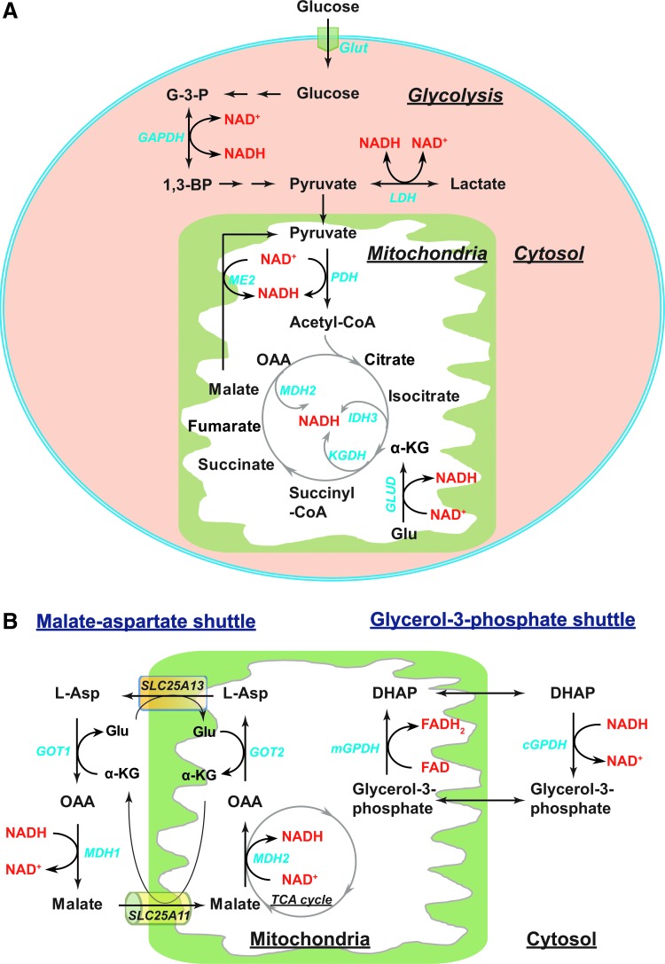
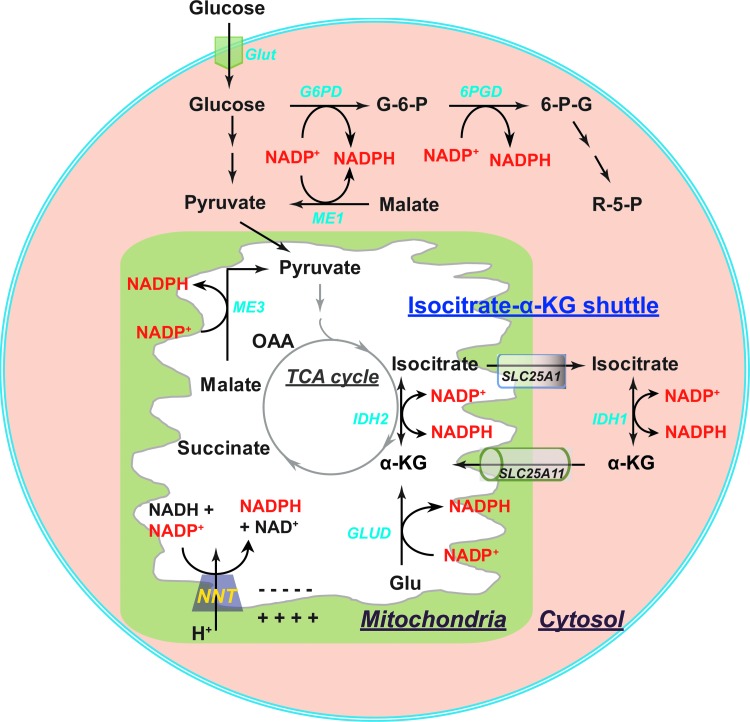
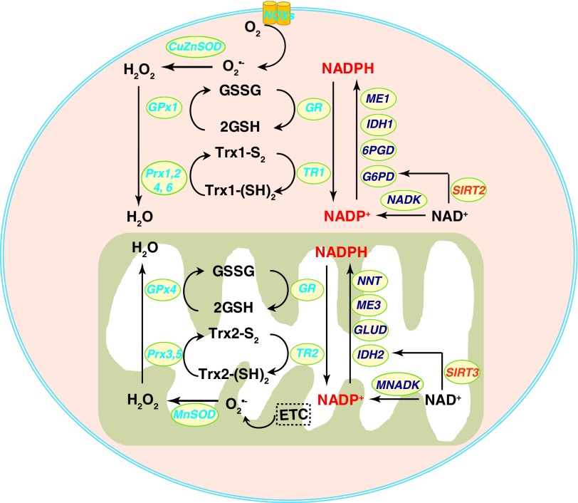
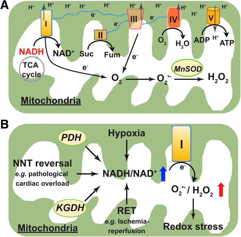
Significance: The nicotinamide adenine dinucleotide (NAD+)/reduced NAD+ (NADH) and NADP+/reduced NADP+ (NADPH) redox couples are essential for maintaining cellular redox homeostasis and for modulating numerous biological events, including cellular metabolism. Deficiency or imbalance of these two redox couples has been associated with many pathological disorders. Recent Advances: Newly identified biosynthetic enzymes and newly developed genetically encoded biosensors enable us to understand better how cells maintain compartmentalized NAD(H) and NADP(H) pools. The concept of redox stress (oxidative and reductive stress) reflected by changes in NAD(H)/NADP(H) has increasingly gained attention. The emerging roles of NAD+-consuming proteins in regulating cellular redox and metabolic homeostasis are active research topics.

Critical issues: The biosynthesis and distribution of cellular NAD(H) and NADP(H) are highly compartmentalized. It is critical to understand how cells maintain the steady levels of these redox couple pools to ensure their normal functions and simultaneously avoid inducing redox stress. In addition, it is essential to understand how NAD(H)- and NADP(H)-utilizing enzymes interact with other signaling pathways, such as those regulated by hypoxia-inducible factor, to maintain cellular redox homeostasis and energy metabolism.

Future directions: Additional studies are needed to investigate the inter-relationships among compartmentalized NAD(H)/NADP(H) pools and how these two dinucleotide redox couples collaboratively regulate cellular redox states and cellular metabolism under normal and pathological conditions. Furthermore, recent studies suggest the utility of using pharmacological interventions or nutrient-based bioactive NAD+ precursors as therapeutic interventions for metabolic diseases. Thus, a better understanding of the cellular functions of NAD(H) and NADP(H) may facilitate efforts to address a host of pathological disorders effectively. Antioxid. Redox Signal. 28, 251-272.

Keywords: NAD(H); NADP(H); cellular metabolism; oxidative stress; redox state; reductive stress.


Figures

<b>FIG. 1.</b>
FIG. 1.
Schematic structures of NAD+(A), NAD+ precursors, and NAD+ derivatives (B-J).
<b>FIG. 2.</b>
FIG. 2.
Biosynthesis of NAD(P)+ in mammalian cells. NAD+ is synthesized by three pathways: the de novo pathway, the Preiss–Handler pathway, and the salvage pathway. De novo NAD+ synthesis from L-Trp is mediated by enzymes in the kynurenine pathway. IDO/TDO catalyzes the first and rate-limiting step by converting L-Trp to N-formylkynurenine. After four enzymatic reactions, the intermediate ACMS undergoes spontaneous cyclization to form QA, which is the second rate-limiting step. QA is then converted to NAMN by QPRT using PRPP as a cosubstrate. In the Preiss–Handler pathway, NA is first metabolized into NAMN by NAPRT. The de novo and Preiss–Handler pathways converge at NAMN, which is further metabolized into NAAD by three NMNATs at the expense of ATP. NAD+ is synthesized from NAAD under the catalysis of NADSYNs. NAM and NR serve as NAD+ precursors for the salvage pathway, in which NAM and NR are initially converted into a common product, NMN, by NAMPT or NRK, respectively. Following that conversion, NMN is metabolized to NAD+ by the same NMNAT enzymes used by the other two pathways. Once formed, NAD+ can be phosphorylated into NADP+ by NADK. ACMS, 2-amino-3-carboxy-muconate-semialdehyde; IDO, indoleamine 2,3-dioxygenase; K3H, kynurenine-3-hydroxylase; KFase, kynurenine formamidase; NA, nicotinic acid; NAAD, NA adenine dinucleotide; NADK, NAD+ kinase; NADSYN, NAD+ synthetase; NAM, nicotinamide; NAMN, nicotinic acid mononucleotide; NAMPT, NAM phosphoribosyltransferase; NAPRT, NA phosphoribosyltransferase; NMNAT, NMN adenylyltransferase; NR, nicotinamide riboside; NRK, NR kinase; PRPP, phosphoribosyl pyrophosphate; QA, quinolinic acid; QPRT, quinolinate phosphoribosyltransferase; TDO, tryptophan 2,3-dioxygenase; Trp, tryptophan.
<b>FIG. 3.</b>
FIG. 3.
A network of NAD+-related enzymes and proteins derived from String v10. Network nodes represent proteins and edges represent protein–protein functional associations with evidence from different sources, as illustrated in the figure. Those interactions ascertained “from curated databases” and “experimentally determined” are known protein–protein interactions imported from databases of physical interactions and experimental repositories. Those interactions derived from “gene neighborhood,” “gene fusions,” mean that proteins fused in some genomes are very likely to be functionally linked. “Gene co-occurrence,” “gene coexpression,” and “protein homology” denote protein–protein interactions predicted by computational methods using genomic information; “gene neighborhood” assumes that a similar genomic context in different species indicates similar functions of the proteins; “gene fusions” means that proteins fused in some genomes are very likely to be functionally linked; “gene co-occurrence” assumes that proteins with an occurrence in the same pathway tend to have similar functions; “gene coexpression” represents predicted protein–protein associations based on observed patterns of similar expression of genes; and “protein homology” refers to protein–protein association predicted by using homologous relationships. “Text mining” uses a large body of scientific texts to search for statistically relevant co-occurrences of genes. HIF-1α, hypoxia-inducible factor 1α; MNADK, mitochondrial NADK; NRF2, nuclear factor (erythroid-derived 2)-like 2; PARP, poly(ADP)-ribosyl polymerase; SIRT, sirtuin family deacetylase.
<b>FIG. 4.</b>
FIG. 4.
Compartmentalization of cellular NAD(H) and NADP(H). In the extracellular milieu, NAD+ from exogenous sources or exported from cells by connexin 43 (Cx43) hemichannels undergoes a series of sequential reactions forming NAM, NMN, NR by ectoenzyme CD38, eNAMPT, and CD73. While extracellular NAM, NMN, and NA are membrane permeable and freely enter the cytosol, extracellular NR is imported via a nucleoside transporter (NT). Once in the cytosol, these precursors can generate NAD(H) and NADP(H) as illustrated in Figure 2. Exogenous NADH is imported into the cytosol via P2X7 receptor-mediated endocytosis. NAD+ can be consumed by cytosolic and nuclear NAD+-dependent proteins (SIRT1, 2, 6, and 7 as well as PARPs) to form NAM, which feeds into the salvage pathway to synthesize NAD+. In mitochondria, NAD+ is synthesized from NMN by NMNAT3 and can be consumed by mitochondrial proteins, such as SIRT3-5 and PARP1, forming NAM. It is proposed that NAM can be converted into NMN by iNAMPT in mitochondrial compartment. NADP+ is formed by MNADK-catalyzed phosphorylation of NAD+. eNAMPT, extracellular NAMPT; iNAMPT, intracellular NAMPT; NADH, reduced NAD+.
<b>FIG. 5.</b>
FIG. 5.
Metabolic sources of NAD(H) and cytosolic/mitochondrial NADH shuttles. (A) In the cytosol, interconversion of NAD+ and NADH is mediated by the glycolytic enzymes GAPDH and LDH. In the mitochondrial matrix, PDH, ME2, GLUD, and TCA cycle enzymes (IDH3, KGDH, and MDH2) contribute to NAD(H) production. (B) Cytosolic and mitochondrial NADH are exchanged through two shuttles: the malate–aspartate shuttle and the glycerol-3-phosphate shuttle. In the malate–aspartate shuttle, cytosolic MDH1 and mitochondrial MDH2 catalyze the reversible interconversion of OAA and malate in conjunction with the interconversion of NAD+ and NADH. Cytosolic GOT1 and mitochondrial GOT2 catalyze the reversible conversion between OAA and L-Asp coupled with the interconversion of Glu and α-KG. The α-KG/malate antiporter (encoded by SLC25A11 gene) and aspartate-glutamate antiporter (encoded by SLC25A13) transport intermediate metabolites between cytosol and mitochondria. In this shuttle, NADH is oxidized to NAD+ in cytosol and NAD+ is reduced to NADH in mitochondria. In the glycerol-3-phosphate shuttle, cytosolic GPDH reduces the glycolytic intermediate DHAP into glycerol-3-phosphate and simultaneously oxidizes NADH to NAD+ in the cytoplasm. Mitochondrial GPDH catalyzes the reverse reaction by oxidizing glycerol-3-phosphate into DHAP and transferring electrons to FAD forming FADH2. α-KG, α-ketoglutarate; DHAP, dihydroxyacetone phosphate; FAD, flavin-adenine dinucleotide; G3P, glyceraldehyde-3-phosphate; GAPDH, glyceraldehyde phosphate dehydrogenase; Glu, glutamate; GLUD, glutamate dehydrogenases; Glut, glucose transporters; GOT, glutamate-OAA transaminase; GPDH, glycerol-3-phosphate dehydrogenase; IDH, isocitrate dehydrogenase; KGDH, α-ketoglutarate dehydrogenase; LDH, lactate dehydrogenase; MDH, malate dehydrogenase; ME, malic enzyme; OAA, oxaloacetate; PDH, pyruvate dehydrogenase; SCL25A11 and SCL25A13, solute carrier family 25 member 11 and 13, respectively; TCA, tricarboxylic acid.
<b>FIG. 6.</b>
FIG. 6.
Metabolic sources of NADP(H) and the cytosolic/mitochondrial NADPH shuttle. In the cytosol, NADPH is primarily produced by G6PD and 6PGD in the pentose phosphate pathway. ME1 also contributes to cytosolic NADPH production. Mitochondrial NADPH is generated by NADP+-dependent IDH2, GLUD, NNT, and ME3. The cytosolic and mitochondrial NADPH is exchanged through the isocitrate-α-KG shuttle, where cytosolic IDH1 and mitochondrial IDH2 catalyze the interconversion of isocitrate and α-KG in conjunction with the interconversion of NADP+ and NADPH. The citrate carrier protein (encoded by SLC25A1 gene) and the α-KG/malate antiporter (encoded by SLC25A11 gene) mediate the transport of isocitrate and α-KG between cytosol and mitochondria, respectively. 6PG, 6-phosphogluconate; 6PGD, 6-phosphogluconate dehydrogenase; G6P, glucose-6-phosphate; G6PD, glucose-6-phosphate dehydrogenase; NNT, nicotinamide nucleotide transhydrogenase; R5P, ribose-5-phosphate; SCL25A1, solute carrier family 25 member 1.
<b>FIG. 7.</b>
FIG. 7.
NADPH and NAD+ function as cofactors in antioxidant defense systems. NADPH is an essential cofactor of GR and TRs. GR catalyzes the recycling of GSH from GSSG, and TRs reduces oxidized Trx-S2 into Trx-(SH)2. Simultaneously, both enzymes require NADPH as an electron donor and oxidize it to NADP+, which can be reduced back to NADPH by ME1, IDH1, G6PD, and G6PD in the cytoplasm, and NNT, ME3, GLUD, and IDH2 in the mitochondria. Once O2•− is formed, for example, from NOXs in the cytosol and from mitochondrial ETC, cytosolic CuZnSOD and mitochondrial MnSOD reduce it to H2O2. GSH can be used by GPx to reduce H2O2 further to water. Trx-(SH)2 provides reducing equivalents for Prx in the removal of H2O2. NAD+ is required for SIRT deacetylase activity. Cytosolic SIRT2 enhances G6PD activity, and mitochondrial SIRT3 enhances IDH2 activity, to enhance the generation of NADPH from NADP+. CuZnSOD, copper zinc superoxide dismutase; ETC, electron transport chain; GPx, glutathione peroxidase; GR, glutathione reductase; GSH, glutathione; GSSG, oxidized GSH; H2O2, hydrogen peroxide; MnSOD, manganese superoxide dismutase; NOX, NADPH oxidase; Prx, peroxiredoxin; TR, thioredoxin reductase; Trx-S2, oxidized thioredoxin; Trx-(SH)2, reduced thioredoxin.
<b>FIG. 8.</b>
FIG. 8.
NADH as pro-oxidant induces redox stress. (A) ROS production by the mitochondrial ETC under physiological conditions. NADH generated primarily in the TCA cycle is oxidized to NAD+ at mitochondrial respiratory complex I (NADH dehydrogenase). Subsequently, electrons from NADH in conjunction with electrons from the oxidation of succinate at complex II (succinate dehydrogenase) are relayed through the mitochondrial ETC and eventually reduce the oxygen molecule to water at complex IV (cytochrome C oxidase). This process is coupled with pumping protons (H+) from the mitochondrial matrix into the intermembrane space at complex I, III, and IV generating an electrochemical proton gradient, which drives ATP production at complex V (ATP synthase). Under physiological conditions, ∼0.1% to 0.2% of total oxygen consumed gains electrons from mitochondrial complex I and III leakage to form O2•−, which is rapidly converted to the more stable H2O2 by MnSOD or by spontaneous dismutation. A steady level of ROS is beneficial and required for many biological processes. (B) Under stressed states, such as hypoxia, NNT reversal, and RET, the mitochondrial NADH/NAD+ ratio increases leading to complex I dysfunction and ROS production. In addition, the NADH-dependent mitochondrial enzymes KGDH and PDH also contribute to mitochondrial ROS production. Overloaded levels of ROS result in redox stress, which is detrimental to cellular function. Fum, fumarate; PDH, pyruvate dehydrogenase; RET, reverse electron transfer; ROS, reactive oxygen species; Suc, succinate.
<b>FIG. 9.</b>
FIG. 9.
NAD+ regulates cellular metabolism. Intracellular NAD+ levels can be increased by supplementing with NAD+ precursors, enhancing the expression and activity of NAD+ biosynthetic enzymes, or by inhibiting NAD+ consumption by PARPs and CD38 enzymes. Increased NAD+ levels further enhance the activity of SIRT proteins. SIRT1, 3, and 6 are deacetylases, whereas SIRT4 is an ADP-ribosylase. SIRT1 deacetylates and activates FOXO1 and PGC-1 resulting in the stimulation of mitochondrial oxidative phosphorylation (OXPHOS) and FA oxidation. SIRT1 can also deacetylate and activate AceCS1 and HIF-2α promoting FA synthesis and glutaminolysis, respectively. By contrast, SIRT1 and SIRT6 deacetylate and inactivate HIF-1α suppressing glycolysis. Similar to SIRT1, SIRT3 deacetylation can also increase mitochondrial OXPHOS and FA synthesis and oxidation. The mitochondria-localized enzyme, SIRT3, can deacetylate mitochondrial complex I and II proteins to enhance their activity; it also targets and activates AceCS2 and LCAD to enhance FA synthesis and oxidation, respectively. Unlike SIRT1, 3, and 6, SIRT4 ADP-ribosylates and inhibits GLUD activity leading to suppression of insulin secretion under basal and stimulated conditions. AceCS, acetyl-CoA synthetase; FA, fatty acids; FOXO1, forkhead box O1; LCAD, long-chain acyl coenzyme A dehydrogenase; OXPHOS, oxidative phosphorylation; PGC-1, peroxisome proliferator-activated receptor γ coactivator 1.

References

    1. Adam-Vizi V. and Chinopoulos C. Bioenergetics and the formation of mitochondrial reactive oxygen species. Trends Pharmacol Sci 27: 639–645, 2006 - PubMed
    1. Agledal L, Niere M, and Ziegler M. The phosphate makes a difference: Cellular functions of NADP. Redox Rep 15: 2–10, 2010 - PMC - PubMed
    1. Ahn BH, Kim HS, Song S, Lee IH, Liu J, Vassilopoulos A, Deng CX, and Finkel T. A role for the mitochondrial deacetylase Sirt3 in regulating energy homeostasis. Proc Natl Acad Sci U S A 105: 14447–14452, 2008 - PMC - PubMed
    1. Aksoy P, Escande C, White TA, Thompson M, Soares S, Benech JC, and Chini EN. Regulation of SIRT 1 mediated NAD dependent deacetylation: A novel role for the multifunctional enzyme CD38. Biochem Biophys Res Commun 349: 353–359, 2006 - PubMed
    1. Alberati-Giani D, Cesura AM, Broger C, Warren WD, Rover S, and Malherbe P. Cloning and functional expression of human kynurenine 3-monooxygenase. FEBS Lett 410: 407–412, 1997 - PubMed