Review
. 2017 Sep 21;24(9):1181-1190.
doi: 10.1016/j.chembiol.2017.05.024. Epub 2017 Jun 22.

Targeted Protein Degradation: from Chemical Biology to Drug Discovery

Affiliations
Review

Targeted Protein Degradation: from Chemical Biology to Drug Discovery

Philipp M Cromm et al. Cell Chem Biol. .

Abstract

Traditional pharmaceutical drug discovery is almost exclusively focused on directly controlling protein activity to cure diseases. Modulators of protein activity, especially inhibitors, are developed and applied at high concentration to achieve maximal effects. Thereby, reduced bioavailability and off-target effects can hamper compound efficacy. Nucleic acid-based strategies that control protein function by affecting expression have emerged as an alternative. However, metabolic stability and broad bioavailability represent development hurdles that remain to be overcome for these approaches. More recently, utilizing the cell's own protein destruction machinery for selective degradation of essential drivers of human disorders has opened up a new and exciting area of drug discovery. Small-molecule-induced proteolysis of selected substrates offers the potential of reaching beyond the limitations of the current pharmaceutical paradigm to expand the druggable target space.

Keywords: E3 ligase; PROTACs; hydrophobic tagging; protein degradation.


Figures

Figure 1
Figure 1. Pharmacology models
Many diseases are caused by abnormal protein function. Occupancy-driven pharmacology blocks malfunctioning proteins via inhibition, i.e., applying high concentrations of inhibitor. In event-driven pharmacology, protein function is controlled by decreasing the cellular protein level. The disease-implicated protein is displayed in dark yellow, the applied inhibitor in red.
Figure 2
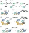
Figure 2. The ubiquitination machinery
(A) ATP dependent ubiquitin activation by ubiquitin-activating enzyme (E1) and transfer to an ubiquitin-conjugating enzyme (E2) results in an E2–Ub intermediate. (B) For RING E3s, ubiquitin is directly linked to the protein of interest (POI) after simultaneous binding to E2–Ub and POI. (C) Simplified schematic blueprint of a Cullin RING E3. Cullin RING E3s are multi-subunit RING ligases comprising an F-box protein for substrate recognition, a RING domain for E2– Ub binding and additional regulatory proteins (omitted for simplicity – more detailed information about Cullin RING ligases can be found elsewhere (Hua and Vierstra, 2011)). (D) HECT- and RBR-type E3s catalyze the ubiquitin transfer in a two-step process. Before ubiquitin is tethered to the POI it is trans-thiolated to the active site of the E3.
Figure 3
Figure 3. Small molecule protein degradation techniques
(A) HyTs degrade their protein target via a not fully elucidated mechanism following one of two possible pathways. (a) HyT binding destabilizes the POI which recruits a chaperone that induces POI proteasomal degradation. (b) The chaperone recognizes the HyT directly and marks the tagged POI for destruction. (B) PROTAC mode of action. The bifunctional PROTAC binds simultaneously to the POI and an E3 bringing both proteins in spatial proximity and inducing ubiquitination. The ubiquitinated POI is subsequently degraded by the proteasome releasing the PROTAC. Both approaches, HyT and PROTACs, can traverse multiple circles allowing for substochiometric usage.
Figure 4
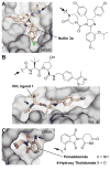
Figure 4. Structural elucidation of E3 ligand binding
(A) Crystal structure of nutlin 3a bound to its target protein MDM2 (PDB: 4HG7). (B) The VHL ligand 1 provides the essential hydroxyproline to engage the F-box protein VHL (PDB: 4W9H). (C) Pomalidomide bound to its molecular target the F-box protein CRBN (PDB: 4TZU). Sites of linker attachment are highlighted by a black arrow.
Figure 5
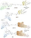
Figure 5
Small molecule PROTACs targeting BET proteins.
Figure 6
Figure 6. Structural insight into PROTAC mode of action
(A) Binding interface between BRD4 (dark grey) and VHL (grey) with MZ1 (light orange) embedded in a bowl-shaped cavity formed by both proteins. The warhead JQ1 as well as E3 recruiting VHL ligand 1 form contacts with both proteins BRD4 and VHL, respectively. (B) Upon formation of the ternary complex BRD4 and VHL engage in extensive PPIs resulting in high cooperativity. (C) Rationally designed BRD4 selective PROTAC AT1. (PDB: 3T35).

References

    1. Adjei AA. What is the right dose? The elusive optimal biologic dose in phase I clinical trials. J Clin Oncol. 2006;24:4054–4055. - PubMed
    1. Bai L, Zhou B, Yang C-Y, Ji J, McEachern D, Przybranowski S, Jiang H, Hu J, Xu F, Zhao Y, et al. Targeted degradation of BET proteins in triple-negative breast cancer. Cancer Res. 2017 doi: 10.1158/0008-5472.CAN-16-2622. - DOI - PMC - PubMed
    1. Bargagna-Mohan P, Baek SH, Lee H, Kim K, Mohan R. Use of PROTACS as molecular probes of angiogenesis. Bioorg Med Chem Lett. 2005;15:2724–2727. - PMC - PubMed
    1. Berndsen CE, Wolberger C. New insights into ubiquitin E3 ligase mechanism. Nat Struct Mol Biol. 2014;21:301–307. - PubMed
    1. Bielskiene K, Bagdoniene L, Mozuraitiene J, Kazbariene B, Janulionis E. E3 ubiquitin ligases as drug targets and prognostic biomarkers in melanoma. Medicina (Kaunas) 2015;51:1–9. - PubMed

MeSH terms