

Current concepts of diagnosis and management of pericardial cysts
- PMID: 28648435
- PMCID: PMC5485391
- DOI: 10.1016/j.ihj.2017.02.021
Current concepts of diagnosis and management of pericardial cysts
Abstract
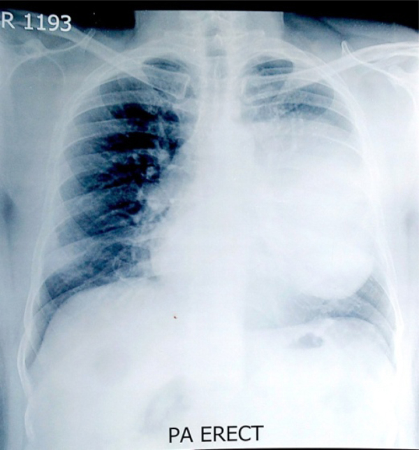
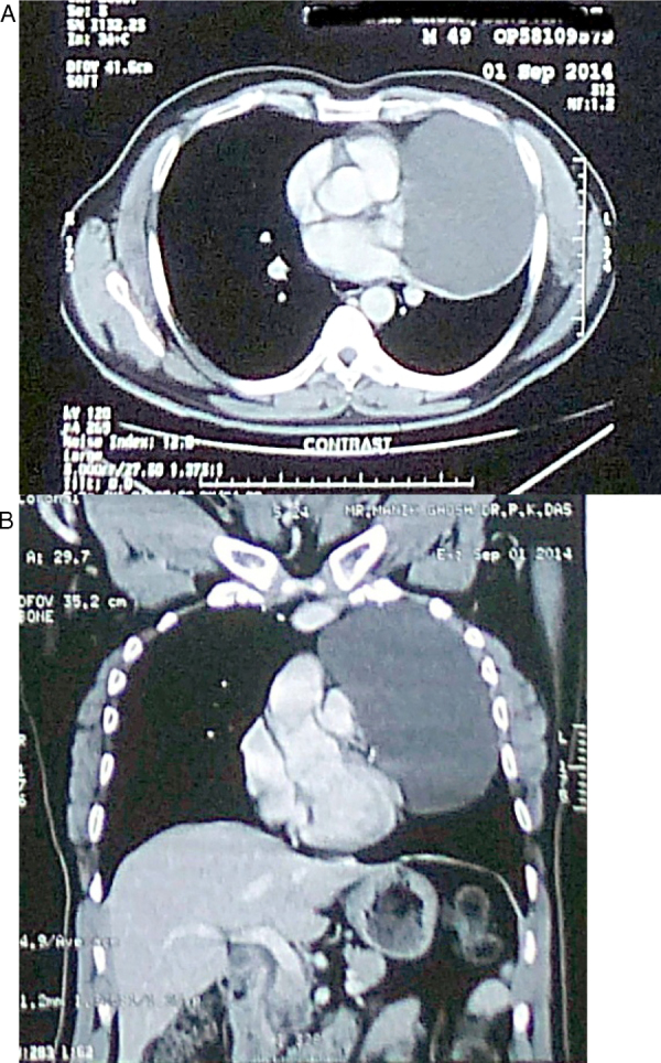
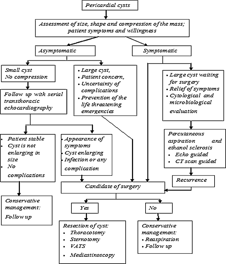
Pericardial cysts are rare with an incidence of about 1 in every 100,000 persons and one in 10 pericardial cysts may actually be a pericardial diverticulum. Pericardial cysts and diverticula share similar developmental origin and may appear as an incidental finding in chest roentgenogram in an asymptomatic patient. CT scan is considered as best modality for diagnosis and delineation of the surrounding anatomy. Cardiac MRI is recommended in the evaluation of the compressive effects caused by the pericardial cysts. The authors recommend echocardiography for serial follow up and image guided aspiration of the pericardial cyst in presence of compressive effects leading to cardiovascular and airway symptoms. A systematic approach is desirable for management of pericardial cysts depending on size, shape and compression effects, symptoms and easy access to serial Echocardiographic follow up. However, pericardial diverticulum may not be differentiated from cysts by the above testing, and only identified at surgery.
Keywords: Algorithmic approach; Historical perspective; Mesothelial cyst; Pericardial coelomic cyst; Pericardial cyst; Pericardial diverticulum; Springwater cyst; Thin walled cyst.
Copyright © 2017. Published by Elsevier B.V.
Figures
References
-
- Davis R.D., Jr., Oldham H.N., Jr., Sabiston D.C., Jr. Primary cysts and neoplasms of the mediastinum: recent changes in clinical presentation, methods of diagnosis, management, and results. Ann Thorac Surg. 1987;44(3):229–237. - PubMed
-
- Cohen A.J., Thompson L., Edwards F.H., Bellamy R.F. Primary cysts and tumors of the mediastinum. Ann Thorac Surg. 1991;51(March (3)):378–384. discussion 385-6. - PubMed
-
- Unverferth D.V., Wooley C.F. The differential diagnosis of paracardiac lesions: pericardial cysts. Cathet Cardiovasc Diagn. 1979;5(1):31–40. - PubMed
-
- Patel J., Park C., Michaels J., Rosen S., Kort S. Pericardial cyst: case reports and a literature review. Echocardiography. 2004;21(April (3)):269–272. - PubMed
Publication types
MeSH terms
LinkOut - more resources
Full Text Sources
Other Literature Sources
Medical
