Review
. 2017 May-Jun;69(3):375-381.
doi: 10.1016/j.ihj.2017.02.015. Epub 2017 Feb 24.

Utility of adjunctive modalities in Coronary chronic total occlusion intervention

Affiliations
Review

Utility of adjunctive modalities in Coronary chronic total occlusion intervention

Hemal Bhatt et al. Indian Heart J. 2017 May-Jun.

Abstract

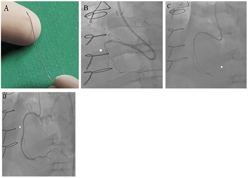
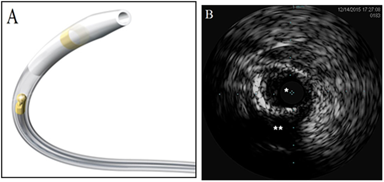
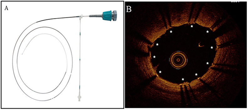
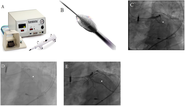
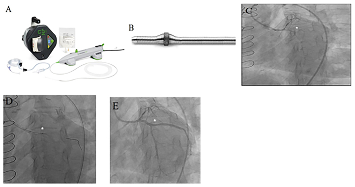
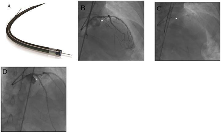
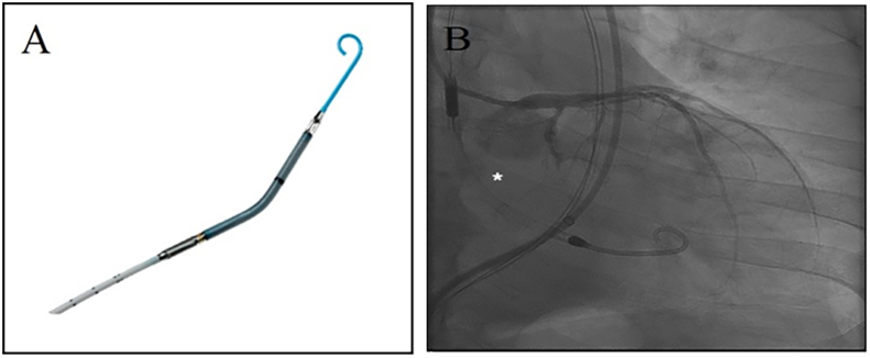
Coronary chronic total occlusion (CTO) intervention remains one of the most challenging domains in interventional cardiology. Due to the technical challenges involved and potential procedural complications, CTO percutaneous coronary intervention (PCI) attempt and success rates remain less than standard PCI. However, the use of several adjunctive tools such as intravascular ultrasound, optical coherence tomography, rotational atherectomy, orbital atherectomy, excimer laser coronary atherectomy and percutaneous left ventricular assist device may contribute to improved CTO PCI success rates or provide better hemodynamic assessment of CTO lesion (i.e., using fractional flow reserve). In this review we present the current literature describing the utility and efficacy of these adjunctive modalities in CTO intervention.

Keywords: Adjunctive modalities; Chronic total occlusion; Fractional flow reserve; Intravascular ultrasound; Percutaneous coronary intervention.


Figures

Fig. 1
Fig. 1
Device and case images of coronary fractional flow reserve measurement (FFR). A. FloWire Doppler Guide Wire for FFR measurement (Image provided courtesy of Volcano Corporation, San Diego, CA); B. Case image demonstrating a proximal right coronary artery (RCA) chronic total occlusion (CTO) (*); C. pre-intervention FFR wire positioning in the distal RCA (*) through a Corsair catheter after crossing the CTO with pre-FFR of 0.45; D. and final post-intervention image of revascularized RCA CTO (*) for post-FFR of 0.85.
Fig. 2
Fig. 2
Device and case images of coronary intravascular ultrasound (IVUS). A. OptiCross IVUS coronary imaging catheter (Image provided courtesy of Boston Scientific Corporation, Natick, MA); B. IVUS case image demonstrating recanalized calcified true lumen (*) and subintimal lumen (**) of chronic total occlusion.
Fig. 3
Fig. 3
Device and case images of coronary optical coherence tomography (OCT). A. Dragonfly duo OCT imaging catheter (Image provided courtesy of St. Jude Medical Inc., Little Canada, MN); B. OCT case image demonstrating adequately expanded and apposed stent struts (*) following PCI of right coronary artery chronic total occlusion.
Fig. 4
Fig. 4
Device and case images of coronary rotational atherectomy (RA). A. RA system and B. Rotablater diamond tipped burr (Images provided courtesy of Boston Scientific Corporation, Natick, Massachusetts); C. Case image demonstrating left circumflex (LCX) chronic total occlusion (CTO) (*); D. Use of RA (*) to treat a heavily calcified segment of LCX CTO [4c]; E. Final angiography of revascularized LCX CTO (*) after RA and stenting.
Fig. 5
Fig. 5
Device and case images of orbital atherectomy (OA). A. Diamondback OA system; B. OA catheter with diamond coated crown (Image provided courtesy of Cardiovascular Systems Inc., St. Paul, MN); C. Case image demonstrating left circumflex (LCX) chronic total occlusion (CTO) (*); D. use of OA (*) in percutaneous coronary intervention of a calcified LCX CTO; E. Final angiography of revascularized LCX CTO (*) after OA and stenting.
Fig. 6
Fig. 6
Device and case images of Excimer laser coronary atherectomy. A. ELCA catheter (Image provided courtesy of Spectranetics, Colorado Springs, CO); B. Case images demonstrating a proximal left anterior descending (LAD) chronic total occlusion (CTO) (*); C. Use of ELCA (*) to debulk the proximal LAD CTO after crossing; D. Final angiography of revascularized LAD CTO (*) after ELCA and stenting.
Fig. 7
Fig. 7
Device and case images of Impella 2.5 LP system. A. Impella 2.5 LP system (Image provided courtesy of Abiomed Corporation, Danvers, MA); B. Case image of percutaneous left ventricular assist device use (*) during percutaneous coronary intervention of left circumflex chronic total occlusion.

References

    1. Shah P.B. Management of coronary chronic total occlusion. Circulation. 2011;123:1780–1784. - PubMed
    1. Bardaji A., Rodriguez-Lopez J., Torres-Sanchez M. Chronic total occlusion: to treat or not to treat. World J Cardiol. 2014;6:621–629. - PMC - PubMed
    1. Aziz S., Ramsdale D.R. Chronic total occlusions–a stiff challenge requiring a major breakthrough: is there light at the end of the tunnel? Heart. 2005;91(Suppl. (3)):42–48. - PMC - PubMed
    1. Grantham J.A., Marso S.P., Spertus J. Chronic total occlusion angioplasty in the United States. JACC Cardiovasc Interv. 2009;2:479–486. - PubMed
    1. Levine G.N., Bates E.R., Blankenship J.C. ACCF/AHA/SCAI guideline for percutaneous coronary intervention: a report of the American College of Cardiology Foundation/American Heart Association task force on practice guidelines and the Society for Cardiovascular Angiography and Interventions. Catheter Cardiovasc Interv. 2011;2013(82):E266–355. - PubMed

MeSH terms