Review
. 2017 Aug;33(8):553-565.
doi: 10.1016/j.tig.2017.05.006. Epub 2017 Jun 22.

Advances in Decoding Axolotl Limb Regeneration

Affiliations
Review

Advances in Decoding Axolotl Limb Regeneration

Brian J Haas et al. Trends Genet. 2017 Aug.

Abstract

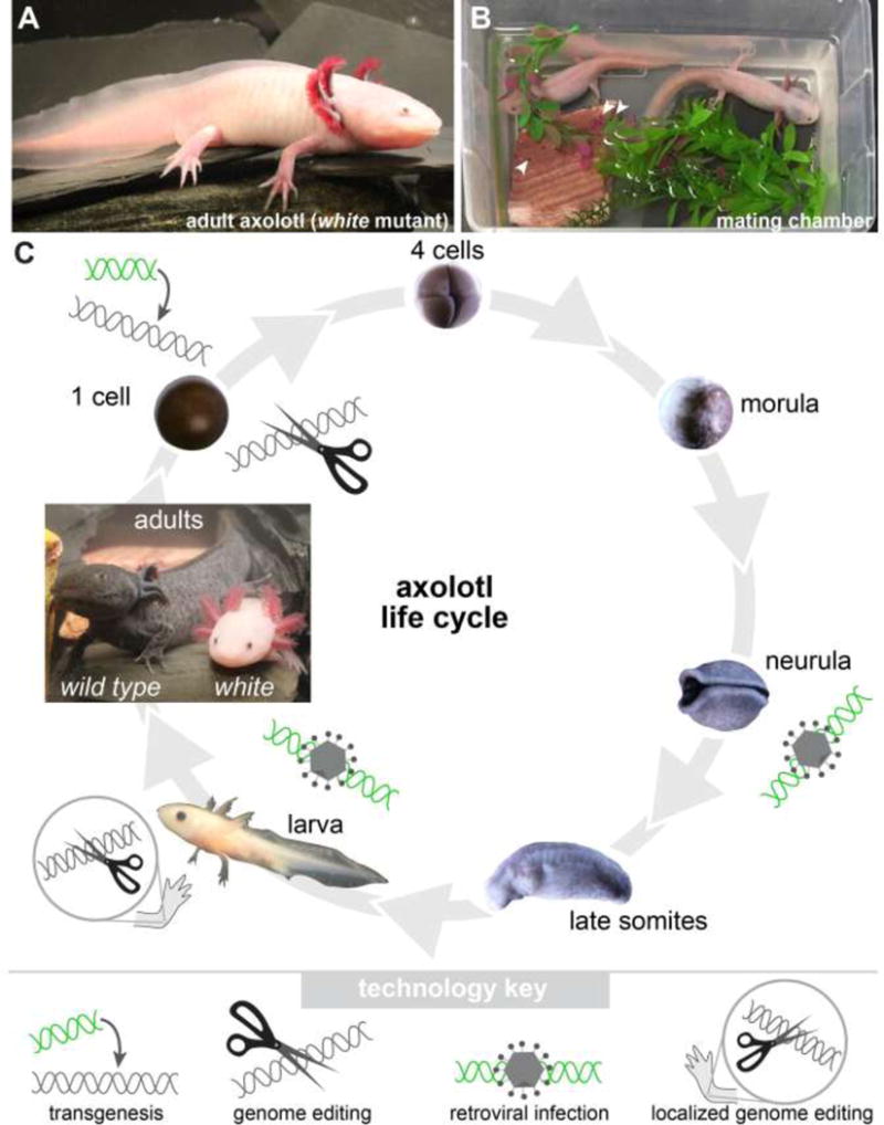
Humans and other mammals are limited in their natural abilities to regenerate lost body parts. By contrast, many salamanders are highly regenerative and can spontaneously replace lost limbs even as adults. Because salamander limbs are anatomically similar to human limbs, knowing how they regenerate should provide important clues for regenerative medicine. Although interest in understanding the mechanics of this process has never wavered, until recently researchers have been vexed by seemingly impenetrable logistics of working with these creatures at a molecular level. Chief among the problems has been the very large size of salamander genomes, and not a single salamander genome has been fully sequenced to date. Recently the enormous gap in sequence information has been bridged by approaches that leverage mRNA as the starting point. Together with functional experimentation, these data are rapidly enabling researchers to finally uncover the molecular mechanisms underpinning the astonishing biological process of limb regeneration.


Figures

Figure 1
Figure 1. Axolotl basics and genome-modifying tools
(A) Shown is an adult axolotl of the white genotype. Note the large limbs. Forelimbs have four digits, while hindlimbs have five digits. (B) Male and female axolotl in mating chamber with eggs. (C) Axoltol life cycle with validated points for genomic modification manipulations noted. Axolotl embryogenesis spans approximately 10–12 days; only some stages are shown. Embryos develop within a transparent jelly coat, which must be removed to permit injections. Two adults are shown. At left is the wild-type genotype whose skin is a darkly-pigmented mottled brownish-black. At right is the white mutant. Note that specimens are not drawn to scale. Techniques that modify the genome are noted at stages where the techniques have been employed to date.
Figure 2
Figure 2. Outline of cellular events during limb regeneration
(A) General progression from unamputated to fully regenerated. (1) Immediately following amputation (within ~24 hours), a thin wound epidermis (magenta) forms across the cut stump via migration of stump epidermal cells. Wound epidermis thickens as cells within it proliferate. (2) A visible bump, called a blastema (blue), forms beneath the wound epidermis. Blastema cells are derived from activated progenitor cells within various stump tissues that migrate to the tip. (3) Blastema cells proliferate to expand the progenitor pool. (4) The initial regeneration response resolves, cells begin to undergo differentiation, and the limb continues to grow to the appropriate size. (B) Early steps are shown in more detail in inset. Architectures of tissues such as bone and muscle are locally deconstructed near the amputation plane and are therefore shown as jagged. Newly-activated progenitor cells, which give rise to future blastema cells, are depicted with bright green starburst outlines. These cells are cued to re-enter the cell cycle and some fraction of them presumably migrate to the space immediately below the wound epidermis. Blood cells, both red and white, are intermingled with blastema cells. A “nascent blastema” is equivalent to very early-bud stage blastema in other literature. Noted are: epidermis (e), wound epidermis (we), dermis (d), bone (b, medium gray), muscle (m, pink), nerves (nv, black). Migration of newly-activated progenitor cells to the tip of the stump during blastema formation is implied by the green arrows.

References

    1. Spallanzani L. Reproductions of the legs in the aquatic salamander., in An Essay on Animal Reproductions [Prodromo di un opera da imprimersi sopra la riproduzioni anamali] Becket and de Hondt; London: 1769. pp. 68–72. [Italian: 1768]
    1. Schreckenberg GM, Jacobson AG. Normal stages of development of the axolotl. Ambystoma mexicanum. Dev Biol. 1975;42(2):391–400. - PubMed
    1. Bordzilovskaya NP, Dettlaff TA, Duhon ST, Malacinski GM. Developmental-stage series of axolotl embryos. In: Armstrong JB, Malacinski GM, editors. Developmental Biology of the Axolotl. Oxford University Press; New York: 1989. pp. 201–219.
    1. Sobkow L, et al. A germline GFP transgenic axolotl and its use to track cell fate: dual origin of the fin mesenchyme during development and the fate of blood cells during regeneration. Dev Biol. 2006;290(2):386–97. - PubMed
    1. Flowers GP, et al. Highly efficient targeted mutagenesis in axolotl using Cas9 RNA-guided nuclease. Development. 2014;141(10):2165–71. - PMC - PubMed

Publication types

LinkOut - more resources