. 2017 Nov;130(Pt B):281-287.
doi: 10.1016/j.pbiomolbio.2017.06.009. Epub 2017 Jun 22.

Impact of titin strain on the cardiac slow force response

Affiliations

Impact of titin strain on the cardiac slow force response

Younss Ait-Mou et al. Prog Biophys Mol Biol. 2017 Nov.

Abstract

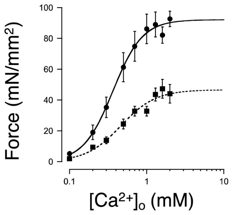
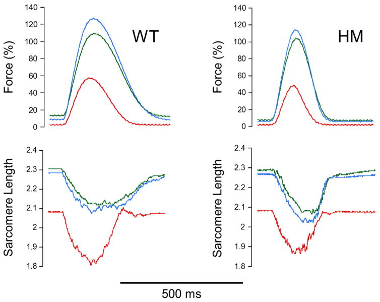
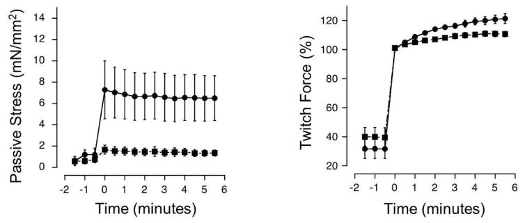
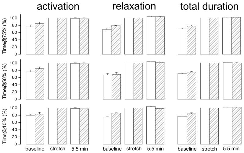
Stretch of myocardium, such as occurs upon increased filling of the cardiac chamber, induces two distinct responses: an immediate increase in twitch force followed by a slower increase in twitch force that develops over the course of several minutes. The immediate response is due, in part, to modulation of myofilament Ca2+ sensitivity by sarcomere length (SL). The slowly developing force response, termed the Slow Force Response (SFR), is caused by a slowly developing increase in intracellular Ca2+ upon sustained stretch. A blunted immediate force response was recently reported for myocardium isolated from homozygous giant titin mutant rats (HM) compared to muscle from wild-type littermates (WT). Here, we examined the impact of titin isoform on the SFR. Right ventricular trabeculae were isolated and mounted in an experimental chamber. SL was measured by laser diffraction. The SFR was recorded in response to a 0.2 μm SL stretch in the presence of [Ca2+]o = 0.4 mM, a bathing concentration reflecting ∼50% of maximum twitch force development at 25 °C. Presence of the giant titin isoform (HM) was associated with a significant reduction in diastolic passive force upon stretch, and ∼50% reduction of the magnitude of the SFR; the rate of SFR development was unaffected. The sustained SL stretch was identical in both muscle groups. Therefore, our data suggest that cytoskeletal strain may underlie directly the cellular mechanisms that lead to the increased intracellular [Ca2+]i that causes the SFR, possibly by involving cardiac myocyte integrin signaling pathways.

Keywords: Isolated cardiac trabeculae; Passive force; Rat; Stretch; Twitch force.


Conflict of interest statement

The authors declare no conflict of interest.

Figures

Figure 1
Figure 1. Average twitch force – extracellular [Ca2+]o relationships
Twitch stress (force divided by cross-sectional area) was measured in electrically stimulated right ventricular trabeculae isolated from wild-type (WT; closed circles; n=7), and giant titin homozygous mutant (HM; closed squares; n=8) rats at bath [Ca2+]o ranging between 0.1 and 2.0 mM. Data were fit to a modified Hill equation (see also Table 1). Presence of the giant isoform in the HM muscles was associated with lower twitch tress (at all [Ca2+]o). In contrast, neither the apparent sensitivity to extracellular Ca2+ (EC50), nor the level of cooperativity (Hill coefficient) was affected by titin isoform (see Table 1). Systolic SL=2.2 μm; stimulus frequency=1 Hz; temperature 25 °C.
Figure 2
Figure 2. Typical Slow Force Response recordings in wild-type (WT) and giant titin homozygous mutant (HM) isolated rat cardiac trabeculae
Sarcomere stretch (ΔSL=0.2 μm) from baseline diastolic SL (red traces) led to an immediate increase in twitch force (green traces); maintaining diastolic SL at the longer length for a further 5.5 minutes induced a further increase in twitch force (blue traces), reflecting the Slow Force Response (SFR) phenomenon (see also Figure 3 and Table 2). The SFR was smaller in HM compared to WT muscles; twitch kinetics were, overall, faster in HM compared to WT muscles, consistent with the average twitch kinetics recorded at saturating bath [Ca2+]o (2 mM; Table 1). [Ca2+]o=0.4 mM; stimulus frequency=1 Hz; temperature=25 °C.
Figure 3
Figure 3. Average passive stress and normalized active twitch force response to stretch
The Slow Force Response (SFR) was recorded in WT (closed circles; n=5) and HM (closed squares; n=8) at [Ca2+]o=0.4 mM (which results in each muscle group ~50% of maximum twitch force). Twitch force was first recorded at the baseline diastolic sarcomere length (SL=2.1 μm) for 1.5 mins; at time=0 min, the muscles were quickly stretched during diastole to increase diastolic sarcomere length to SL=2.3 μm (ΔSL=0.2 μm), resulting in an immediate increase in both passive (left panel) and active twitch force (right panel). In the right panel, active twitch force is normalized to peak twitch force of the first elicited twitch following the stretch. Presence of the giant isoform of titin in the HM muscles was associated with significant lower passive stress development following stretch and a ~50% lower final magnitude of the SFR in HM muscles. The kinetics of the SFR, however, were not affected (see also Table 2). [Ca2+]o=0.4 mM; stimulus frequency=1 Hz; temperature=25 °C.
Figure 4
Figure 4. Impact of stretch on twitch kinetics
Average twitch kinetics at the 75% (top), 50% (middle) and 10% (bottom) twitch force level at: baseline sarcomere length (left pair of bars), immediately following stretch (middle pair of bars), and 5.5 min following the stretch when the Slow Force Response is fully developed (right pair of bars). Data were recorded in wild-type (WT; open bars; n=5) and homozygous giant titin mutant (HM; hatched bars; n=8) muscles. Time durations were normalized to the first twitch immediately following the stretch (i.e. the middle pair of bars representing the immediate response to SL stretch was set to 100%). In both muscle groups and at all three force levels, stretch per se induced an increase in time to reach peak twitch force (activation, left panels) and between the peak of the contraction down to a given twitch force level (relaxation, middle panels), and the overall duration of the twitch (total duration, right panels); prolonged maintained stretch (5.5 minutes), in contrast, did not affect twitch kinetics. [Ca2+]o=0.4 mM; temperature=25 °C; stimulus frequency=1.0 Hz.

References

    1. Ait Mou Y, Hsu K, Farman GP, Kumar M, Greaser ML, Irving TC, de Tombe P. Titin strain contributes to the Frank-Starling law of the heart by structural rearrangements of both thin- and thick-filament proteins. Proc Natl Acad Sci USA. 2016;113:2306–2311. - PMC - PubMed
    1. Ait Mou Y, le Guennec J-Y, Mosca E, de Tombe PP, Cazorla O. Differential contribution of cardiac sarcomeric proteins in the myofibrillar force response to stretch. Pflugers Arch. 2008;457:25–36. - PMC - PubMed
    1. Allen DG, Kurihara S. The effects of muscle length on intracellular calcium transients in mammalian cardiac muscle. J Physiol. 1982;327:79–94. - PMC - PubMed
    1. Anrep von G. On the part played by the suprarenals in the normal vascular reactions of the body. J Physiol. 1912;45:307–317. - PMC - PubMed
    1. Cingolani HE, Ennis IL, Aiello EA, Pérez NG. Role of autocrine/paracrine mechanisms in response to myocardial strain. Pflugers Arch. 2011;462:29–38. - PubMed

Publication types