. 2017 Sep:162:9-17.
doi: 10.1016/j.exer.2017.06.016. Epub 2017 Jun 23.

EphA2 and ephrin-A5 are not a receptor-ligand pair in the ocular lens

Affiliations

EphA2 and ephrin-A5 are not a receptor-ligand pair in the ocular lens

Catherine Cheng et al. Exp Eye Res. 2017 Sep.

Abstract

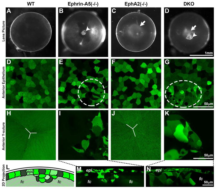
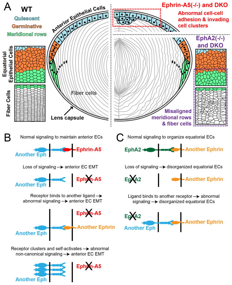
Eph-ephrin bidirectional signaling is essential for eye lens transparency in humans and mice. Our previous studies in mouse lenses demonstrate that ephrin-A5 is mainly expressed in the anterior epithelium, where it is required for maintaining the anterior epithelial monolayer. In contrast, EphA2 is localized in equatorial epithelial and fiber cells where it is essential for equatorial epithelial and fiber cell organization and hexagonal cell shape. Immunostaining of lens epithelial and fiber cells reveals that EphA2 and ephrin-A5 are also co-expressed in anterior fiber cell tips, equatorial epithelial cells and newly formed lens fibers, although they are not precisely colocalized. Due to this complex expression pattern and the promiscuous interactions between Eph receptors and ephrin ligands, as well as their complex bidirectional signaling pathways, cataracts in ephrin-A5(-/-) or EphA2(-/-) lenses may arise from loss of function or abnormal signaling mechanisms. To test whether abnormal signaling mechanisms may play a role in cataractogenesis in ephrin-A5(-/-) or EphA2(-/-) lenses, we generated EphA2 and ephrin-A5 double knockout (DKO) mice. We compared the phenotypes of EphA2(-/-) and ephrin-A5(-/-) lenses to that of DKO lenses. DKO lenses displayed an additive lens phenotype that was not significantly different from the two single KO lens phenotypes. Similar to ephrin-A5(-/-) lenses, DKO lenses had abnormal anterior epithelial cells leading to a large mass of epithelial cells that invade into the underlying fiber cell layer, directly resulting in anterior cataracts in ephrin-A5(-/-) and DKO lenses. Yet, similar to EphA2(-/-) lenses, DKO lenses also had abnormal packing of equatorial epithelial cells with disorganized meridional rows, lack of a lens fulcrum and disrupted fiber cells. The DKO lens phenotype rules out abnormal signaling by EphA2 in ephrin-A5(-/-) lenses or by ephrin-A5 in EphA2(-/-) lenses as possible cataract mechanisms. Thus, these results indicate that EphA2 and ephrin-A5 do not form a lens receptor-ligand pair, and that EphA2 and ephrin-A5 have other binding partners in the lens to help align differentiating equatorial epithelial cells or maintain the anterior epithelium, respectively.


Figures

Fig. 1
Fig. 1
Immunostaining of anterior and equatorial epithelial and fiber cells from lens capsule flat mounts of P21 wild-type lenses for ephrin-A5 (red) and EphA2 (green). (A) Single optical sections (XY views) show that anterior epithelial cells have punctate ephrin-A5 staining signals and no EphA2 staining. In contrast, anterior fiber cells have EphA2 staining along the cell membrane with a few ephrin-A5 puncta. Scale bar, 10 μm. (B) 3D reconstruction of z-stacks (XZ view) shows ephrin-A5 punctate signals are concentrated near the basal and lateral cell membranes of anterior epithelial cells (EC) with some staining also at the apical junction between anterior epithelial cells and the tips of elongating fiber cells (F, arrows). EphA2 is predominately expressed in lens fiber cells. This staining pattern in anterior epithelial and fiber cells is consistent with our previously published data (Cheng and Gong, 2011). (C) Single optical sections show that ephrin-A5 and EphA2 are both present in equatorial epithelial cells organized into meridional rows and in the newly added secondary fiber cells. The EphA2 staining pattern is consistent with our previous results (Cheng et al., 2013). Scale bar, 10 μm. D) 3D reconstruction of z-stacks (YZ view) showing the bow region of the lens reveals the EphA2 and ephrin-A5 staining signals are present in equatorial epithelial cells (EC) and newly formed fibers (F). Arrows point to ephrin-A5 puncta in lens fiber cells.
Fig. 2
Fig. 2
(A) Cartoon of ephrin-A5 (red) and EphA2 (green) localization in lens epithelial and fiber cells. Not drawn to scale. (B) There are three possible mechanisms for cataracts in ephrin-A5(−/−) lenses. Loss of ephrin-A5 leads to EMT in anterior epithelial cells (EC) due to disruption of bidirectional signaling (loss of function). It would be unlikely in this loss of function mechanism for EphA2 to be the receptor due to the divergent phenotypes between EphA2(−/−) and ephrin-A5(−/−) lenses. Alternatively, the ephrin-A5(−/−) lens phenotype might be a consequence of abnormal signaling due to EphA2 binding to a different ligand or self-activating through non-canonical autophosphorylation. (C) In EphA2(−/−) lenses, disorganization of equatorial epithelial cells (EC) may be caused by loss of function. In the loss of function mechanism, ephrin-A5 is unlikely to be the ligand interacting with EphA2 because of the vast differences in EphA2(−/−) versus ephrin-A5(−/−) lens phenotypes. However, it is possible that in the absence of EphA2, ephrin-A5 interacts with another receptor causing abnormal signaling in equatorial epithelial cells.
Fig. 3
Fig. 3
(A–D) Photos of P21 WT, ephrin-A5(−/−), EphA2(−/−) and double knockout (DKO) lenses. Ephrin-A5(−/−) and DKO lenses often have anterior cataracts (B and D, arrowheads). EphA2(−/−) and DKO lenses often display mild nuclear opacities (C and D, arrows). Scale bar, 1mm. (D–G) Confocal images of anterior epithelial cells in P21 GFP+ WT, ephrin-A5(−/−), EphA2(−/−) and DKO lenses. WT and EphA2(−/−) lenses have normal anterior epithelium with cobblestone shaped cells and mosaic GFP expression (D and F). Ephrin-A5(−/−) and DKO lenses display abnormal epithelial cell morphology (E and G, dashed ellipses). Scale bar, 50μm. (H–K) Images of the anterior Y-suture region just beneath the epithelium. WT and EphA2(−/−) lenses have normal Y-shaped sutures (H and J, white lines). The aberrant cluster of epithelial cells in ephrin-A5(−/−) and DKO lenses extends into the anterior suture region (I and K). Scale bar, 50μm. (L) A cartoon depicting a 2D projection of the 3D reconstruction of a Z-stack through the anterior epithelium and underlying fiber cells (not drawn to scale). (M–N) Aberrant clusters of epithelial cells invading the anterior suture and fiber cell layer can be seen in the ephrin-A5(−/−) and DKO lens. Scale bar, 50μm. H–J: epi, epithelial cells; fc, fiber cells.
Fig. 4
Fig. 4
(A–D) Confocal images of equatorial epithelial cells from P21 GFP+ WT, ephrin-A5(−/−), EphA2(−/−) and DKO lenses. WT and ephrin-A5(−/−) lenses display hexagonal equatorial epithelial cells (A and B) aligned into meridional rows (below the white dashed line). In contrast, equatorial epithelial cells in EphA2(−/−) and DKO lenses lack organized meridional rows (C and D). Scale bar, 50μm. (E–H) Confocal images of peripheral fiber cells in P21 GFP+ WT, ephrin-A5(−/−), EphA2(−/−) and DKO lenses. While WT and ephrin-A5(−/−) lenses have straight and organized peripheral differentiating fiber cells (E and F), fiber cells are wavy and disorganized in EphA2(−/−) and DKO lenses (G and H). Scale bar, 50μm. (I–L) Whole mount staining of P21 lenses with DAPI (nuclei) and images from the lens equator. WT and ephrin-A5(−/−) equatorial epithelial cells have organized and aligned rows of nuclei in meridional rows (I and J, arrowheads). However, loss of EphA2 in single KO and DKO lenses leads to disorganized cell nuclei (K and L, arrows) and disrupted meridional rows. Scale bar, 25μm. (M–P) WGA staining of cell membranes in whole mount P21 WT, ephrin-A5(−/−), EphA2(−/−) and DKO lenses. Imaging at the lens equator near the cortex reveals that the lens fulcrum is strongly stained by WGA in WT and ephrin-A5(−/−) lenses (M and N, arrows). In contrast, Epha2(−/−) and DKO lenses lack a well-defined lens fulcrum (O and P). Scale bar, 25μm.
Fig. 5
Fig. 5
(A) A summary of the ephrin-A5(−/−), EphA2(−/−) and DKO lens phenotypes. (Left panel) Normal mouse lenses consist of a monolayer of epithelial cells and bulk elongated fiber cells, wrapped by the lens capsule. Anterior epithelial cells (blue) are quiescent. Equatorial epithelial cells undergo proliferation (orange) and transform from a random cell packing organization into meridional rows of hexagonally packed cells (green). Hexagonal fiber cells retain organized rows. (Right panel) Loss of ephrin-A5 in single knockout and DKO lenses leads to abnormal cell-cell adhesion and clusters of anterior epithelial cells that invade into the underlying fiber cell layer (red box). In contrast, EphA2(−/−) and DKO lenses have disorganized meridional rows and fiber cells at the lens equator (purple box). (B) In the absence of ephrin-A5, dysfunction and EMT in anterior epithelial cells (EC) may result from loss of function or abnormal signaling mechanism through an unknown Eph receptor. (C) In EphA2(−/−) lenses, disorganization of equatorial epithelial cells (EC) may result from loss of function or abnormal ligand signaling.

References

    1. Arvanitis D, Davy A. Eph/ephrin signaling: networks. Genes Dev. 2008;22:416–429. - PMC - PubMed
    1. Asbell PA, Dualan I, Mindel J, Brocks D, Ahmad M, Epstein S. Age-related cataract. Lancet. 2005;365:599–609. - PubMed
    1. Astin JW, Batson J, Kadir S, Charlet J, Persad RA, Gillatt D, Oxley JD, Nobes CD. Competition amongst Eph receptors regulates contact inhibition of locomotion and invasiveness in prostate cancer cells. Nature cell biology. 2010;12:1194–1204. - PubMed
    1. Banh A, Deschamps PA, Gauldie J, Overbeek PA, Sivak JG, West-Mays JA. Lens-specific expression of TGF-beta induces anterior subcapsular cataract formation in the absence of Smad3. Investigative ophthalmology & visual science. 2006;47:3450–3460. - PMC - PubMed
    1. Barquilla A, Lamberto I, Noberini R, Heynen-Genel S, Brill LM, Pasquale EB. Protein kinase A can block EphA2 receptor-mediated cell repulsion by increasing EphA2 S897 phosphorylation. Mol Biol Cell. 2016;27:2757–2770. - PMC - PubMed

Publication types