Review
. 2018 Apr;1864(4 Pt B):1262-1269.
doi: 10.1016/j.bbadis.2017.06.017. Epub 2017 Jun 23.

Mechanisms of cholangiocyte responses to injury

Affiliations
Review

Mechanisms of cholangiocyte responses to injury

Keisaku Sato et al. Biochim Biophys Acta Mol Basis Dis. 2018 Apr.

Abstract

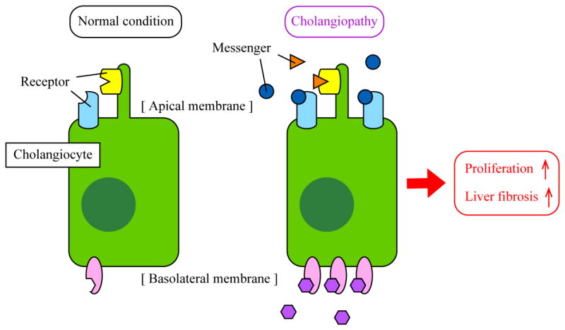
Cholangiocytes, epithelial cells that line the biliary epithelium, are the primary target cells for cholangiopathies including primary sclerosing cholangitis and primary biliary cholangitis. Quiescent cholangiocytes respond to biliary damage and acquire an activated neuroendocrine phenotype to maintain the homeostasis of the liver. The typical response of cholangiocytes is proliferation leading to bile duct hyperplasia, which is a characteristic of cholestatic liver diseases. Current studies have identified various signaling pathways that are associated with cholangiocyte proliferation/loss and liver fibrosis in cholangiopathies using human samples and rodent models. Although recent studies have demonstrated that extracellular vesicles and microRNAs could be mediators that regulate these messenger/receptor axes, further studies are required to confirm their roles. This review summarizes current studies of biliary response and cholangiocyte proliferation during cholestatic liver injury with particular emphasis on the secretin/secretin receptor axis. This article is part of a Special Issue entitled: Cholangiocytes in Health and Diseaseedited by Jesus Banales, Marco Marzioni, Nicholas LaRusso and Peter Jansen.

Keywords: Bile ducts; Biliary damage; Cholangiocytes; Ductular reactions.


Figures

Figure 1
Figure 1. Mechanisms of cholangiocyte proliferation
Small and large cholangiocytes have different mechanisms for proliferation. Proliferation for small cholangiocytes, which are smaller in size and have a larger nucleus to cytosol ratio compared to large cholangiocytes, is dependent on intracellular Ca2+ release induced by IP3 while large cholangiocyte proliferation is cAMP-dependent. Binding of messengers such as histamine for small cholangiocytes and SCT for large cholangiocytes to their corresponding receptors triggers these pathways leading to cholangiocyte proliferation. When large cholangiocytes are damaged by triggers such as γ–aminobutyric acid or CCl4, small cholangiocytes begin to express SR and differentiate into large cholangiocytes in order to maintain bile duct homeostasis and function. Signaling mechanisms on how damaged large cholangiocytes initiate differentiation in small cholangiocytes are still unknown.
Figure 2
Figure 2. The function of the SCT/SR axis in liver diseases
SCT binds to SR that is expressed only in large cholangiocytes in the liver, and SCT binding and SR activation lead to elevated exocytosis and ductular secretion. Recent studies have shown that human patients with cholangiopathies such as PSC show elevated expression of SCT and SR indicating an association between the SCT/SR axis and cholestatic liver injury. Studies using SCT−/− and SR−/− mice have demonstrated that bile duct hyperplasia and liver fibrosis are attenuated in those knockout mice during BDL suggesting the functional contribution of the SCT/SR axis to the pathogenesis of cholangiopathies. A recent study has also demonstrated that the SCT/SR axis may be responsible for liver steatosis leading to NAFLD and NASH.
Figure 3
Figure 3. Working model of cholangiocyte responses during cholangiopathies
Cholangiocytes are quiescent in healthy conditions (left). During cholestatic liver injury, expression levels of messengers such as SCT and SP as well as corresponding receptors such as SR and NK-1R are elevated in cholangiocytes (right). Binding of messengers to receptors in an autocrine/paracrine manner triggers signaling pathways for proliferation and fibrogenesis leading to bile duct hyperplasia and liver fibrosis. Although specific or primary locations are not identified for all proteins, some proteins such as TGR5 are located on primary cilia or apical membrane, and some proteins such as SR and CFTR are located on the basolateral membrane.

References

    1. Maroni L, Haibo B, Ray D, Zhou T, Wan Y, Meng F, Marzioni M, Alpini G. Functional and structural features of cholangiocytes in health and disease. Cell Mol Gastroenterol Hepatol. 2015;1:368–380. - PMC - PubMed
    1. Kanno N, LeSage G, Glaser S, Alpini G. Regulation of cholangiocyte bicarbonate secretion. Am J Physiol Gastrointest Liver Physiol. 2001;281:G612–625. - PubMed
    1. Tabibian JH, Masyuk AI, Masyuk TV, O’Hara SP, LaRusso NF. Physiology of cholangiocytes. Compr Physiol. 2013;3:541–565. - PMC - PubMed
    1. Lesage G, Glaser SS, Gubba S, Robertson WE, Phinizy JL, Lasater J, Rodgers RE, Alpini G. Regrowth of the rat biliary tree after 70% partial hepatectomy is coupled to increased secretin-induced ductal secretion. Gastroenterology. 1996;111:1633–1644. - PubMed
    1. Alvaro D, Mancino MG, Glaser S, Gaudio E, Marzioni M, Francis H, Alpini G. Proliferating cholangiocytes: a neuroendocrine compartment in the diseased liver. Gastroenterology. 2007;132:415–431. - PubMed

MeSH terms