Review
. 2017 Jul 5;26(1):39-48.
doi: 10.1016/j.cmet.2017.05.016. Epub 2017 Jun 22.

Mitochondrial Dynamics in Regulating the Unique Phenotypes of Cancer and Stem Cells

Affiliations
Review

Mitochondrial Dynamics in Regulating the Unique Phenotypes of Cancer and Stem Cells

Hsiuchen Chen et al. Cell Metab. .

Abstract

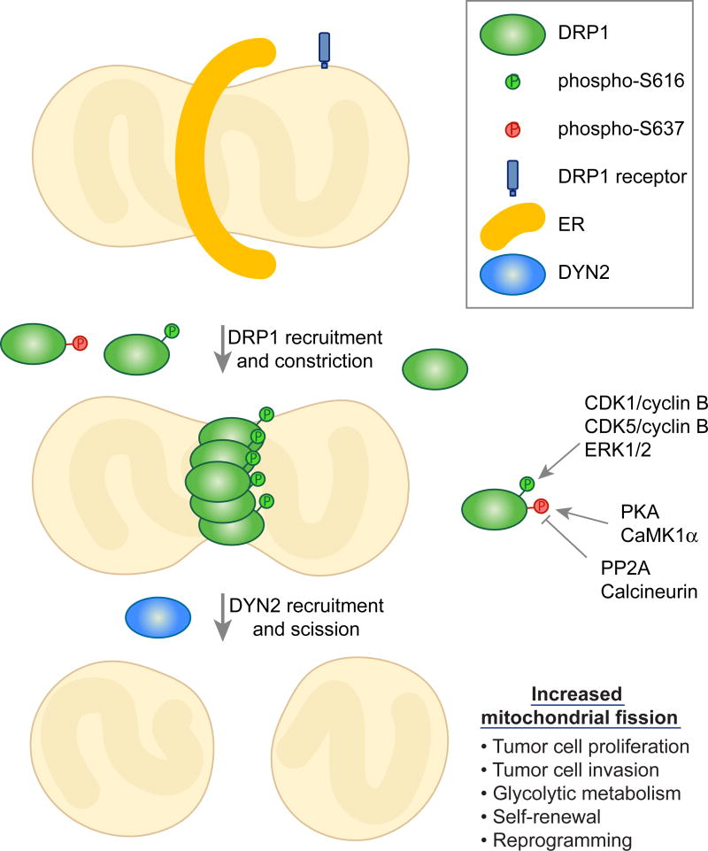
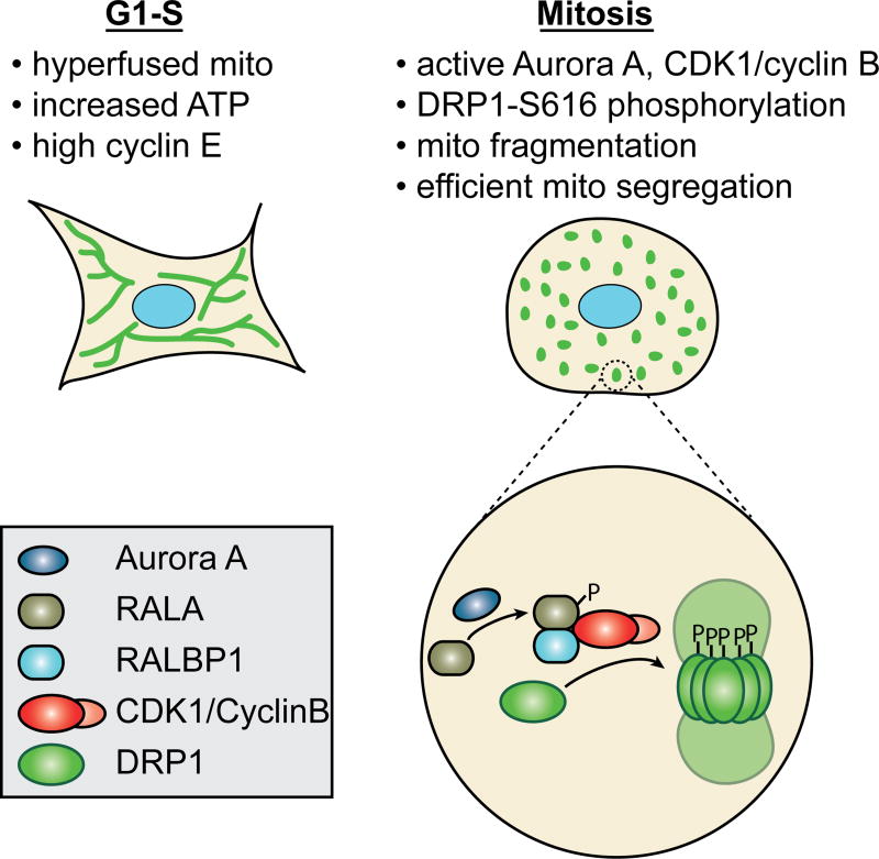
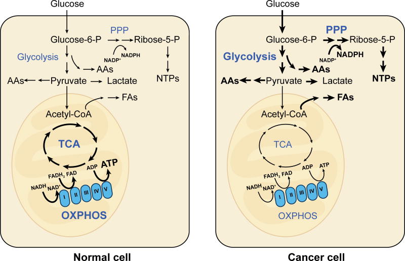
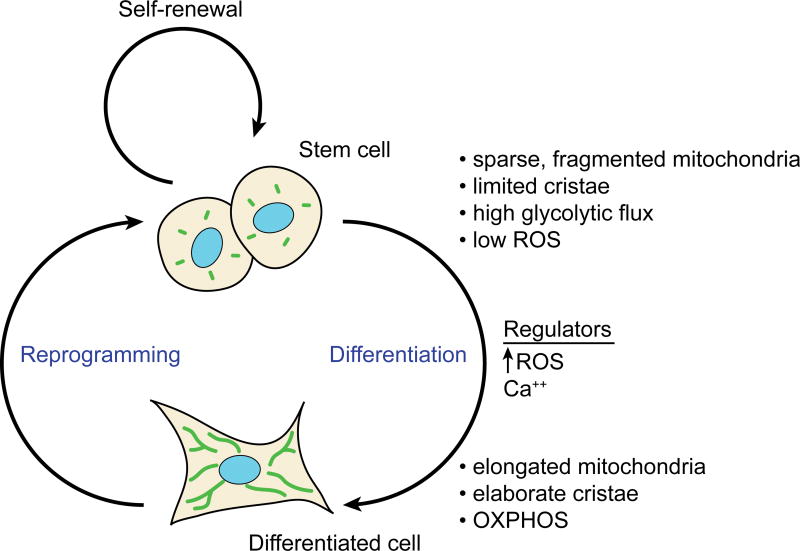
Cancer and stem cells appear to share a common metabolic profile that is characterized by high utilization of glucose through aerobic glycolysis. In the presence of sufficient nutrients, this metabolic strategy provides sufficient cellular ATP while additionally providing important metabolites necessary for the biosynthetic demands of continuous cell proliferation. Recent studies indicate that this metabolic profile is dependent on genes that regulate the fusion and fission of mitochondria. High levels of mitochondrial fission activity are associated with high proliferation and invasiveness in some cancer cells and with self-renewal and resistance to differentiation in some stem cells. These observations reveal new ways in which mitochondria regulate cell physiology, through their effects on metabolism and cell signaling.

Keywords: cancer; induced pluripotential stem cells; metabolism; mitochondria; mitochondrial dynamics; stem cells.


Figures

Figure 1
Figure 1. Regulation of mitochondrial fission and its role in cancer and stem cells
The fission of mitochondria begins with an interaction with the endoplasmic reticulum, which causes initial constriction of the mitochondrial tubule. DRP1, recruited by one of several DRP1 receptors, assembles on the mitochondrial surface and causes further constriction. The final stage of membrane scission requires DYN2. The activity of DRP1 is regulated by molecules that control the phosphorylation of DRP1 at two serine residues, which have opposing effects. The level of mitochondrial fission has significant effects on tumor and stem cell phenotypes.
Figure 2
Figure 2. Mitochondrial dynamics during the cell cycle
The two signature features of mitochondrial structure during the cell cycle are the hyperfused network at G1-S and the extensively fragmented state at mitosis. The hyperfused network at G1-S is associated with increased ATP and high cyclin E levels. The fragmented state during mitosis facilitates equitable distribution of mitochondria to daughter cells. Fragmentation is driven by phosphorylation of DRP1 by CDK1/cyclin B, causing the activation of DRP1 and mitochondrial fission. Aurora A works upstream to activate CDK1/cyclin B.
Figure 3
Figure 3. Metabolic rewiring in normal versus cancer cells
Differentiated cells typically rely heavily on the OXPHOS activity of mitochondria. In contrast, many cancer cells show the Warburg effect, characterized by reliance on aerobic glycolysis and reduced emphasis on OXPHOS. Glycolysis, though less energy efficient than OXPHOS, generates metabolic intermediates that provide building blocks for synthesis of amino acids (AAs), fatty acids (FAs), and nucleotides (NTPs).
Figure 4
Figure 4. Mitochondrial and metabolic profiles of stem cells versus differentiated cells
ESCs have less reliance on mitochondrial metabolism, and this is reflected in the ultrastructure of their mitochondria. Similar differences exist when somatic cells are reprogrammed into iPSCs. As noted in the main text, these generalizations for mitochondrial structure in stem cells do not apply to neural stem cells. ROS and calcium are two regulators of differentiation that are regulated by mitochondrial function.

References

    1. Amati-Bonneau P, Valentino ML, Reynier P, Gallardo ME, Bornstein B, Boissiere A, Campos Y, Rivera H, de la Aleja JG, Carroccia R, et al. OPA1 mutations induce mitochondrial DNA instability and optic atrophy 'plus' phenotypes. Brain. 2008;131:338–351. - PubMed
    1. Bleazard W, McCaffery JM, King EJ, Bale S, Mozdy A, Tieu Q, Nunnari J, Shaw JM. The dynamin-related GTPase Dnm1 regulates mitochondrial fission in yeast. Nat Cell Biol. 1999;1:298–304. - PMC - PubMed
    1. Bordt EA, Clerc P, Roelofs BA, Saladino AJ, Tretter L, Adam-Vizi V, Cherok E, Khalil A, Yadava N, Ge SX, et al. The Putative Drp1 Inhibitor mdivi-1 Is a Reversible Mitochondrial Complex I Inhibitor that Modulates Reactive Oxygen Species. Developmental cell. 2017;40:583–594.e586. - PMC - PubMed
    1. Carelli V, Chan DC. Mitochondrial DNA: impacting central and peripheral nervous systems. Neuron. 2014;84:1126–1142. - PMC - PubMed
    1. Cassidy-Stone A, Chipuk JE, Ingerman E, Song C, Yoo C, Kuwana T, Kurth MJ, Shaw JT, Hinshaw JE, Green DR, et al. Chemical inhibition of the mitochondrial division dynamin reveals its role in Bax/Bak-dependent mitochondrial outer membrane permeabilization. Developmental cell. 2008;14:193–204. - PMC - PubMed