. 2017 Jan:111:1-26.
doi: 10.1016/j.mser.2016.11.001.

Injectable scaffolds: Preparation and application in dental and craniofacial regeneration

Affiliations

Injectable scaffolds: Preparation and application in dental and craniofacial regeneration

Bei Chang et al. Mater Sci Eng R Rep. 2017 Jan.

Abstract

Injectable scaffolds are appealing for tissue regeneration because they offer many advantages over pre-formed scaffolds. This article provides a comprehensive review of the injectable scaffolds currently being investigated for dental and craniofacial tissue regeneration. First, we provide an overview of injectable scaffolding materials, including natural, synthetic, and composite biomaterials. Next, we discuss a variety of characteristic parameters and gelation mechanisms of the injectable scaffolds. The advanced injectable scaffolding systems developed in recent years are then illustrated. Furthermore, we summarize the applications of the injectable scaffolds for the regeneration of dental and craniofacial tissues that include pulp, dentin, periodontal ligament, temporomandibular joint, and alveolar bone. Finally, our perspectives on the injectable scaffolds for dental and craniofacial tissue regeneration are offered as signposts for the future advancement of this field.

Keywords: craniofacial; dental; injectable; scaffold; tissue regeneration.


Figures

Fig. 1
Fig. 1
Chemical structures of representative injectable natural and synthetic biomaterials
Fig. 2
Fig. 2
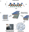
A Self-assembled peptide amphiphile (PA). (a) Chemical structure of the peptide amphiphile. (b) Molecular model of the PA. (c) Schematic showing the self-assembly of PA molecules into a cylindrical micelle. Adapted with permission from [63], Copyright 2001 American Association for the Advancement of Science.
Fig. 3
Fig. 3
Physically crosslinked hydrogels. (a) Ionic crosslinked hydrogels. (b) Hydrogel bond crosslinked hydrogels. (c) Temperature-induced crosslinking hydrogels.
Fig. 4
Fig. 4
A injectable gelatin derivative hydrogel with sustained VEGF release for induced angiogenesis. (a) Synthesis of injectable gelatin-derived hydrogels. (b) Control of the mechanical strength of the gelatin-derived hydrogel using different H2O2 concentrations. (c) The released VEGF from the gelatin-derived hydrogel showed high bioactivity using a chick chorioallantoic membrane (CAM) assay. Adapted with permission from [153]. Copyright 2015 Elsevier Ltd.
Fig. 5
Fig. 5
Scheme of cell-instructive hydrogel preparation using a selective conjugate addition approach. Adapted with permission from [126].Copyright 2003 Macmillan Publishers Ltd.
Fig. 6
Fig. 6
An example of self-assembling peptide hydrogels. (a) The molecular model of SPG-178 peptide, Facio was used: cyan, carbon; red, oxygen; blue, nitrogen; white, hydrogen. (b) A schematic diagram of the formation of the hydrogel from the peptide monomer. Adapted with permission from [153]. Copyright 2011 Elsevier Ltd.
Fig. 7
Fig. 7
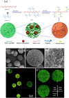
Development of hierarchical nanofibrous microspheres with controlled growth factor delivery for tissue regeneration. (a) Schematic illustration of the hierarchical microsphere structure. (b–f) Characterizations of the hierarchical growth factor-loading microspheres. To visualize the nanospheres and microspheres, TRITC-conjugated gelatin blended with heparin-modified gelatin was used and FITC-conjugated bovine serum albumin (BSA) was adsorbed on the nanofibers of the microspheres. Adapted with permission from [166]. Copyright 2015 WILEY-VCH Verlag GmbH & Co.
Fig. 8
Fig. 8
Regeneration of full-length pulp using hierarchical injectable microspheres as cell carriers. (a) Overview of an SEM image that shows the dental pulp stem cells (DPSCs) adhering to the surfaces of the hierarchical microspheres, expanded their processes and grasped the microspheres to form cell/microsphere aggregates. (b) A typical SEM image of the DPSCs adhered to a microsphere surface, showing the close interaction between the cell and the microsphere. (c) The full-length root canal model. (d) Regenerated pulp-like tissue fulfilled both the apical and middle third regions and reached the coronal third of the canal. A large number of blood vessels were observed throughout the canals. In some regions, DPSCs differentiated into odontoblast-like cells and aligned with the existing tubular dentin of the root. Adapted with permission from [187]. Copyright 2016 Elsevier Ltd.
Fig. 9
Fig. 9
Periodontal tissue regeneration using chitosan-based injectable thermosensitive hydrogel (CS-HTCC/GP) in a dog periodontal defect model. (a) Histological examination of periodontal tissue regeneration in different groups (Mallory's trichrome staining methods). NB, new bone; NC, new cementum; PDL, new periodontal ligaments; SF, Sharpey's fibers; CV, capillary vessel. (b) Height comparision of regenerated periodontal tissues in three groups. Adapted with permission from [160]. Copyright 2010 Elsevier Ltd.
Fig. 10
Fig. 10
Bone regeneration in defect areas around dental implants. (a) Bone regeneration and integration evaluation from histologic images in each group. The red lines indicate the lower boundary of the defect area. F, fibrous tissue; NB, newly formed bone; OB, old bone. (b) Bone remodeling evaluation using fluorescent labelling. Green: calcein at week 2, and red:alizarin reds at week 6.Adapted with permission from [202]. Copyright 2015 IOP Publishing Ltd.
Fig. 11
Fig. 11
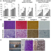
Articular cartilage generation applying PEG-LA-DM/PEGDM copolymer hydrogels. (a) Biochemical evaluation of PEG-LA-DM/PEGDM copolymer/chondrocyte constructs, including DNA content, GAG content and hydroxyproline content. (b) Histological and an immunohistochemical results from a 60/40 ratio of degradable/nondegradable PEG construct compared to native swine articular cartilage. (c) Macroscopic view, toluidine blue staining and collagen type II staining of the integration interface between the engineered cartilage (EC) and native articular cartilage (NC) of 18 week 60/40 construct of ring model for integration study.Adapted from [215]. Open access. Copyright 2016 BioMed Central.

References

    1. Dye BA, Tan S, Smith V, Lewis BG, Barker LK, Thornton-Evans G, Eke PI, Beltran-Aguilar ED, Horowitz AM, Li CH. Vital. Health. Stat. 2007;11:1–92. - PubMed
    1. Eke PI, Dye BA, Wei L, Thornton-Evans GO, Genco RJ. J. Dent. Res. 2012;91:914–920. - PubMed
    1. Adell R, Eriksson B, Lekholm U, Branemark PI, Jemt T. Int. J. Oral. Maxillofac. Implants. 1990;5:347–359. - PubMed
    1. Allen KD, Athanasiou KA. Tissue Eng. 2006;12:1183–1196. - PubMed
    1. Qu T, Jing J, Jiang Y, Taylor RJ, Feng J, Geiger B, Liu X. Tissue Eng. Part A. 2014;17–18:2422–2433. - PMC - PubMed