

Leishmaniasis: a review
- PMID: 28649370
- PMCID: PMC5464238
- DOI: 10.12688/f1000research.11120.1
Leishmaniasis: a review
Abstract
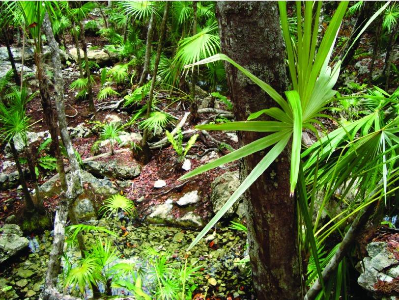
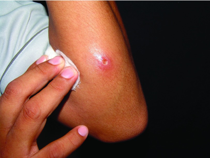
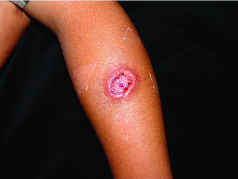
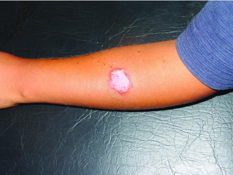
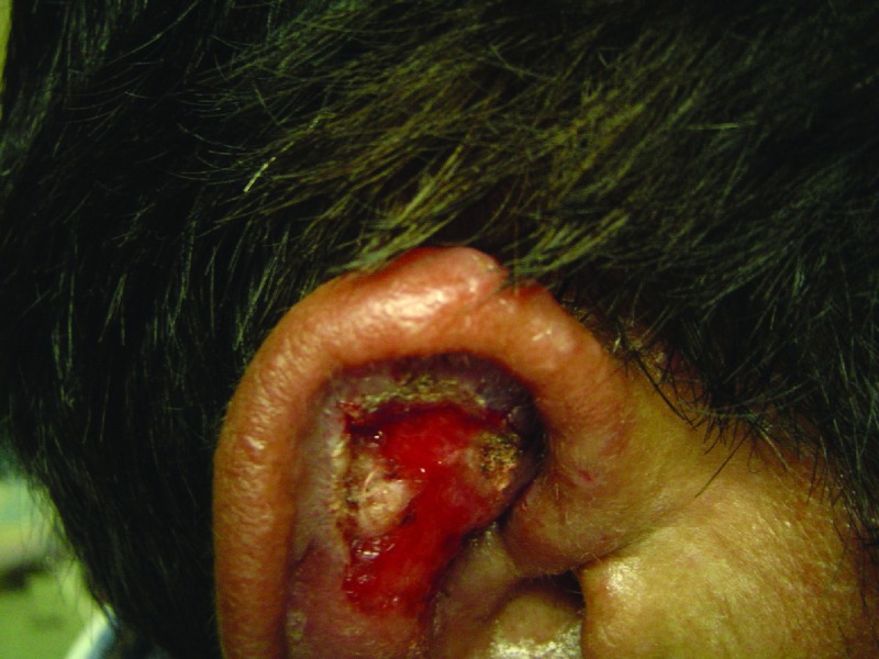
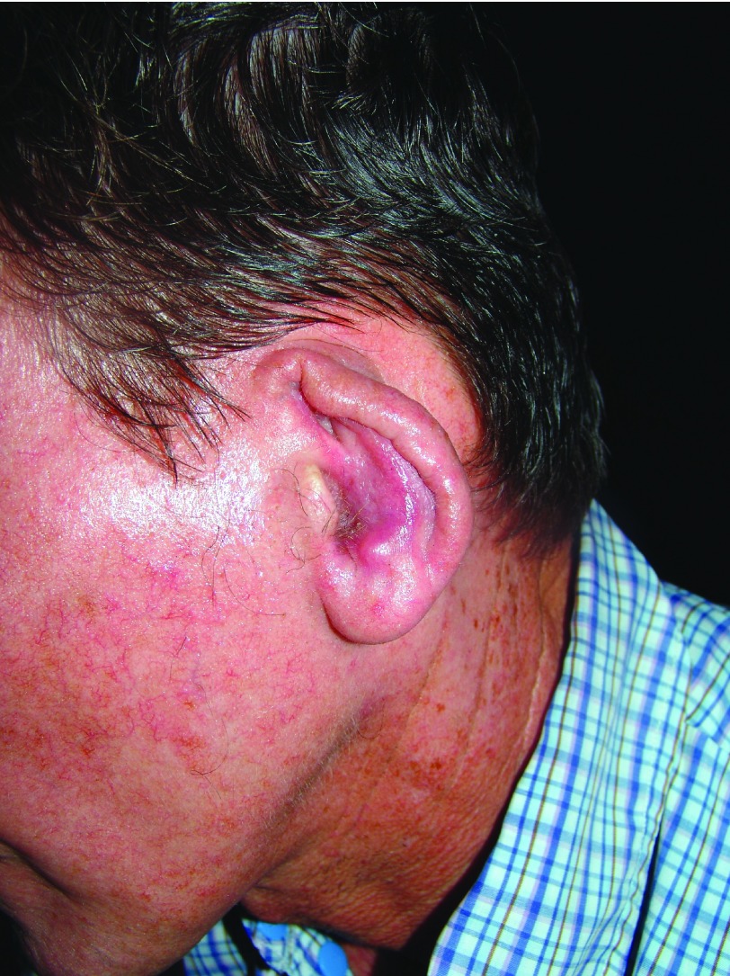
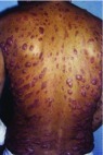
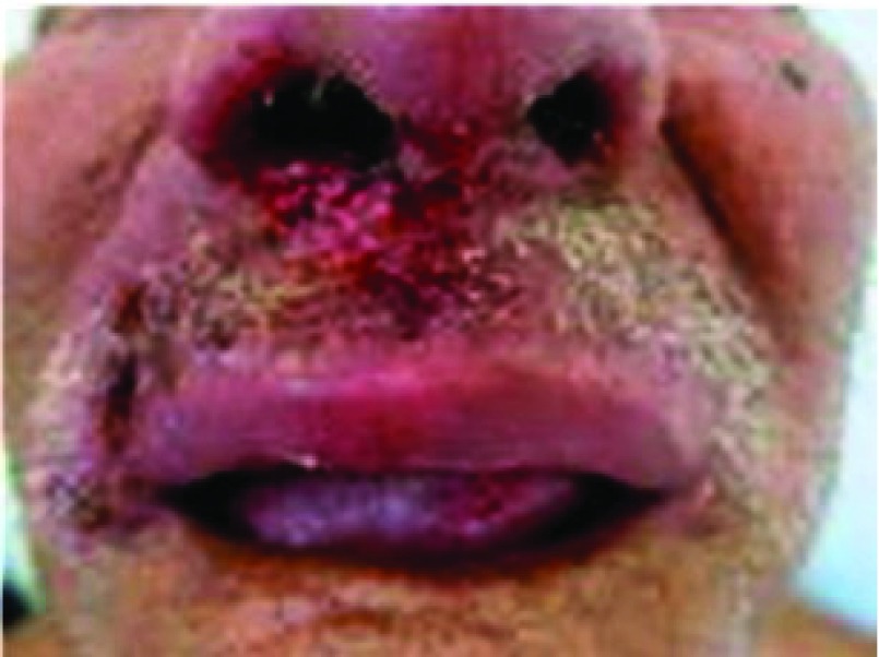
Leishmaniasis is caused by an intracellular parasite transmitted to humans by the bite of a sand fly. It is endemic in Asia, Africa, the Americas, and the Mediterranean region. Worldwide, 1.5 to 2 million new cases occur each year, 350 million are at risk of acquiring the disease, and leishmaniasis causes 70,000 deaths per year. Clinical features depend on the species of Leishmania involved and the immune response of the host. Manifestations range from the localized cutaneous to the visceral form with potentially fatal outcomes. Many drugs are used in its treatment, but the only effective treatment is achieved with current pentavalent antimonials.
Keywords: Leishmania; Leishmaniasis; chicleros ulcer; cutaneous-chondral.
Conflict of interest statement
Competing interests: The authors declare that they have no competing interests.No competing interests were disclosed.No competing interests were disclosed.
Figures
References
-
- Vera-Izaguirre D, Vega-Memije E, Quintanilla Cedillo M, et al. : Leishmaniasis revisión. DCMQ. 2006;4(4):252–260. Reference Source
-
- Vargas-Martínez F, Torres-Guerrero E, Quintanilla-Cedillo MR, et al. : Leishmaniasis en México. Academia Mexicana de Dermatología, Colegio de Dermatólogos de Yucatán A. C., Fundación Mexicana para la Dermatología, Universidad Autónoma de Campeche y Secretaría de Salud, México.2013.
Publication types
LinkOut - more resources
Full Text Sources
Other Literature Sources