. 2017 Apr 25:3:11.
doi: 10.1038/s41522-017-0018-1. eCollection 2017.

Surface topology affects wetting behavior of Bacillus subtilis biofilms

Affiliations

Surface topology affects wetting behavior of Bacillus subtilis biofilms

Moritz Werb et al. NPJ Biofilms Microbiomes. .

Abstract

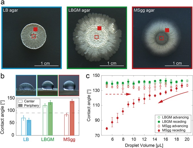
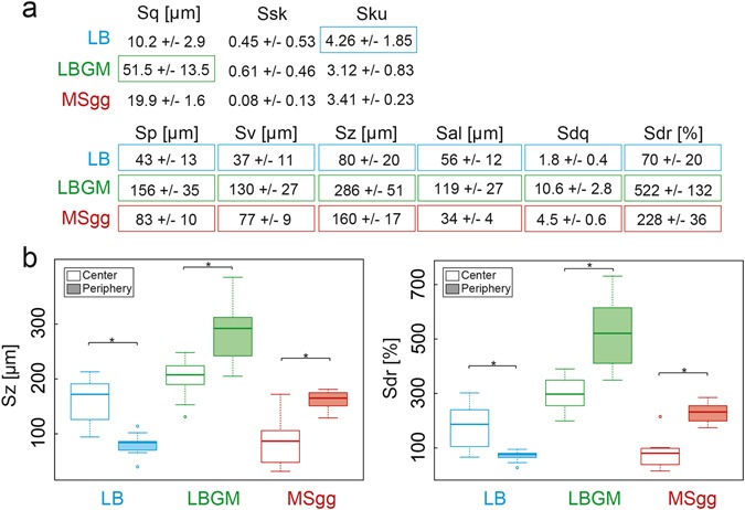
The colonization of surfaces by bacterial biofilms constitutes a huge problem in healthcare and industry. When attempting biofilm inactivation or removal, it is crucial to sufficiently wet the biofilm surface with antibacterial agents; however, certain biofilms efficiently resist wetting, and the origin of this behavior remains to date unclear. Here, we demonstrate that, depending on the growth medium used, the model bacterium Bacillus subtilis can form biofilm colonies with distinct surface properties: we find either hydrophilic or two variants of hydrophobic behavior. We show that those differences in biofilm wetting correlate with distinct surface topologies which, in turn, give rise to different physical wetting regimes known from lotus leaves or rose petals. Forming biofilms with different wetting properties may help bacteria to survive in both arid and humid conditions. Furthermore, converting the surface polarity of a biofilm could facilitate their removal from surfaces by increasing their wettability.


Conflict of interest statement

The authors declare no competing interests.

Figures

Fig. 1
Fig. 1
The wetting behavior of B. subtilis NCIB 3610 biofilms depends on the biofilm growth medium and on the location on the biofilm colony. When B. subtilis NCIB 3610 is grown on LB agar enriched with different molecules, the morphology of the formed macrocolonies changes a and a different wetting behavior of the biofilms is observed (b). In the images shown in a, the regions on the biofilm surface where the wetting tests were performed are marked with a closed and open red square, respectively. The droplet images shown in b were acquired on the peripheral regions of the biofilm colonies. For the peripheral regions of biofilms grown on MSgg agar, a pronounced contact angle hysteresis is observed, but not for biofilms grown on LBGM agar (c). The experimental time scale for the wetting/dewetting experiment was identical for both biofilm variants. Error bars denote the standard deviation. For data shown in b, n ≥ 9; for data shown in c, n = 3
Fig. 2
Fig. 2
The surface topology of B. subtilis NCIB 3610 biofilms depends on the biofilm growth medium. When analyzed by light profilometry, B. subtilis NCIB 3610 biofilms show differences in their height features (a). Similar differences can also be observed in SEM images (b). Furthermore, for biofilms grown on MSgg, the topology in the center and the periphery of the colonies seems to be different
Fig. 3
Fig. 3
Metrological quantification of the surface topologies of B. subtilis NCIB 3610 biofilms. A broad range of metrological parameters a are calculated from the surface profiles obtained from the peripheral regions of biofilms. With a subset of those metrological parameters, both the central and peripheral regions of a given biofilm colony are compared (b). Boxed values in a and asterisks in b indicate statistically significant differences (n ≥ 9, p < 0.05)
Fig. 4
Fig. 4
Surface analysis of the peripheral region of biofilms before, during, and after wetting. Confocal fluorescence images a show differences in biofilm staining after local wetting with a dying solution. Light profilometry images before, during, and after wetting b suggest a homogenously wetted surface for LB biofilms, a three-phase Cassie–Baxter wetting regime for LBGM biofilms, and an impregnated Cassie wetting regime for MSgg biofilms (see main text for details). Please note the much higher resolution of the images in z-direction than in x-direction and y-direction
Fig. 5
Fig. 5
Proteomics analysis of the center and periphery of B. subtilis NCIB 3610 biofilms grown on different agar substrates. For statistical evaluation of the biofilm composition, volcano plots are generated from data of three independent experimental replicates to illustrate differences in protein expression between the center and periphery of the different biofilm types, i.e., LB, LBGM, and MSgg biofilm. The y-axis represents the p-value and the x-axis lists the binary logarithm of the n-fold change in protein expression levels between the center and the periphery of a biofilm colony. The solid lines indicate a significance level of p = 0.05 and a required minimum fold change of 2 (s0 = 1) which is used as a cut-off for significance. The dots below the cut off lines correspond to proteins expressed both in the center and periphery without significant differences. The red dots above the cut off lines represent proteins which are expressed at significantly higher or lower levels in the center compared to the periphery of that colony

References

    1. Neinhuis C, Barthlott W. Characterization and distribution of water-repellent, self-cleaning plant surfaces. Ann. Bot. 1997;79:667–677. doi: 10.1006/anbo.1997.0400. - DOI
    1. Feng L, et al. Super-hydrophobic surfaces: from natural to artificial. Adv. Mater. 2002;14:1857–1860. doi: 10.1002/adma.200290020. - DOI
    1. Bhushan B, Nosonovsky M. The rose petal effect and the modes of superhydrophobicity. Philos. Trans. R. Soc. Lond. A. 2010;368:4713–4728. doi: 10.1098/rsta.2010.0203. - DOI - PubMed
    1. Xu, Q. et al. Robust self-cleaning and micromanipulation capabilities of gecko spatulae and their bio-mimics. Nat. Commun. 6, 8949 (2015). - PMC - PubMed
    1. Gao X, Jiang L. Biophysics: water-repellent legs of water striders. Nature. 2004;432:36–36. doi: 10.1038/432036a. - DOI - PubMed

LinkOut - more resources