

doi: 10.1038/s41551-016-0008.
Epub 2017 Jan 10.
Light in diagnosis, therapy and surgery
Affiliations
- PMID: 28649464
- PMCID: PMC5476943
- DOI: 10.1038/s41551-016-0008
Item in Clipboard
Light in diagnosis, therapy and surgery
Nat Biomed Eng.
2017.
Abstract
Light and optical techniques have made profound impacts on modern medicine, with numerous lasers and optical devices being currently used in clinical practice to assess health and treat disease. Recent advances in biomedical optics have enabled increasingly sophisticated technologies - in particular those that integrate photonics with nanotechnology, biomaterials and genetic engineering. In this Review, we revisit the fundamentals of light-matter interactions, describe the applications of light in imaging, diagnosis, therapy and surgery, overview their clinical use, and discuss the promise of emerging light-based technologies.
Conflict of interest statement
Competing financial interests The author declares no competing financial interests
Figures
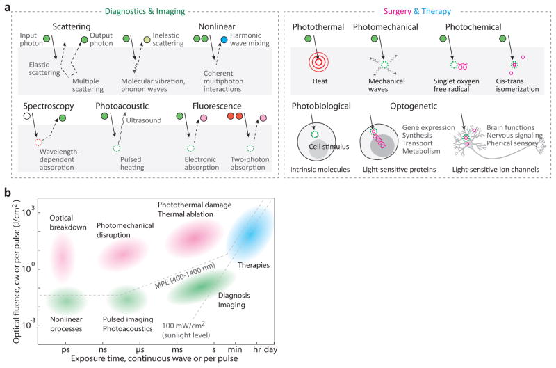
Light–tissue interactions. a, Representative optical
mechanisms used in diagnosis and imaging (left), and surgery and therapy
(right). Circular objects denote incoming and outgoing photons, with their
trajectories indicated by solid and dotted lines with arrowheads. Circle colours
represent the spectrum of the light; dotted circles indicate the absorption of
input photons. For the case of therapy, specific effects of light on tissue and
cells are indicated. b, Optical techniques mapped according to
their optical fluence and exposure time (either total illumination time for cw
light, or pulse duration for pulses). Background colours represent medical
areas: green for diagnostics, magenta for surgery, and cyanine for therapy. MPE,
maximum permissible exposure.
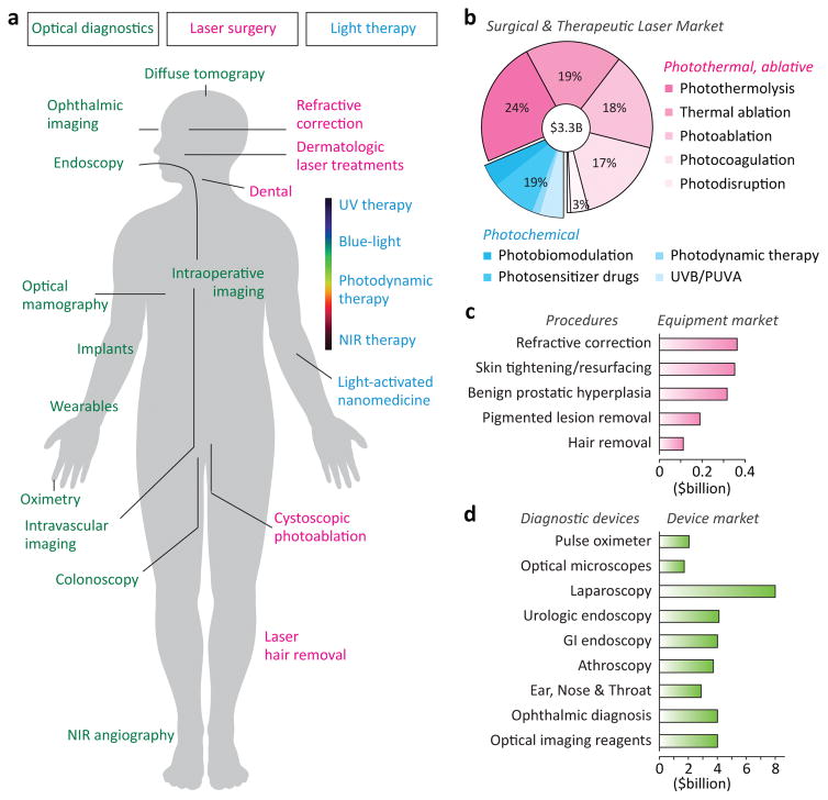
Medical application areas of light. a, Representative applications
of light in the human body for diagnosis and imaging (green), surgery (magenta)
and therapy (cyanine). b, Global market in 2014 for medical lasers
by therapeutic application. Photosensitizer drugs are included as part of the
PDT market. c, Equipment spending in 2013 for leading
laser-treatment procedures. d, Global market in 2013–2014
for several major diagnostic devices. Data sources: BCC Research reports
HLC093C, HLC072C, and HLC172A (with permission of BCC Research, Wesley, MA,
USA).
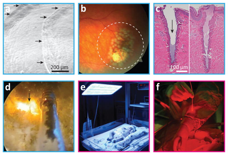
Surgical and therapeutic applications of light. a, Photoablation.
The scanning electron micrograph shows stepwise ablated patterns (arrows) in an
experimental cornea by excimer laser irradiation. b, Photocoagulation.
Laser-induced damage spots around a retinal tear region (dashed-line circle). c,
Photothermal ablation. Histology of porcine skin tissues harvested 0 min (left)
and 60 min (right) after fractional, pulsed CO2 laser exposure
in vivo. The arrow indicates the laser channel, which is filled with
fibrin plug within minutes. d, Photothermal ablation
(vaporization). A fibre-optic catheter delivers high-power continuous-wave laser
(arrow) to remove excess prostate tissue in a patient with benign prostate
hyperplasia [from Feldman, RG. Prostata laser verde green light.
www.youtube.com/watch?v=jAbSwtSN9xE . License at http://creativecommons.org/license/by/3.0 ].
e, Blue-light therapy for the treatment of neonatal jaundice.
f, Photodynamic therapy. Close-up image of a surgeon’s
hands in an operating room. Panel e reproduced with permission from
Photobiological Sciences Online. Panel f courtesy of the National
Cancer Institute.
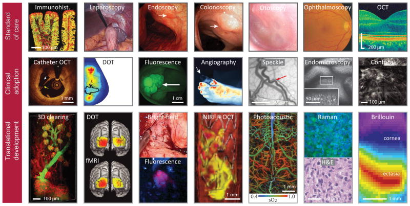
Current and emerging optical imaging. Top row, Established microscopy and
endoscopy in routine clinical use. Immunohistochemistry shows c-Met expression
in an adenomatous polyp.
Laparoscopy visualizes the peritoneal cavity for minimally invasive
surgery. Endoscopy
reveals a mucosal lesion (arrow) in gastric body. Colonoscopy shows a large, 3-cm
sigmoid polyp (arrow).
Otoscopy visualizes the eardrum and middle ear. Ophthalmoscopy fundus camera image of
the human retina. Optical
coherence tomography (OCT) allows microscopic cross-sectional imaging of the
retina. Middle
row, Imaging technologies currently in clinical adoption phases. Diffuse optical
tomography (DOT) shows oxygen saturation map for a breast with a 2.5-cm
malignant tumor (arrow).
Speckle contrast image reveals meningeal artery (arrow) in the cortex and
dura. Fluorescence
image shows colorectal polyps labelled with fluorescent peptides. Fluorescence angiography
shows loss of blood perfusion (arrow) in the foot. Catheter OCT shows plaque rupture in a
human coronary artery, identified by a broken fibrous cap (arrow). Confocal laser endomicroscopy
shows intramucosal bacteria (bright spots, inset) in the small intestine of a
patient with ulcerative colitis. Reflectance confocal microscopy shows an aggregate of
neoplastic cells appearing as a focus of bright nuclei in the
dermal–epidermal junction. Bottom row, Technologies under development or in
clinical testing. Microscopy of optically cleared thick-tissue blocks holds
promise for the high-content mapping and phenotyping of normal and pathological
tissue samples. Functional
connectivity maps of three sensory-motor networks generated with high-density
DOT and fMRI (ref ).
Intraoperative imaging of gliomas under white-light and fluorescence imaging
with 5-aminolevulinic acid. Multimodal (OCT plus fluorescence) image shows a
three-dimensional rendering of the stented right iliac artery of a living
rabbit. Red,
artery wall; white, stent; purple, thrombus; yellow, NIR fluorescent fibrin.
Photoacoustic image of oxygen saturation of haemoglobin (sO2) in the
murine brain vasculature. Clinically viable stimulated Raman microscopy shows
hypercellularity and nuclear atypia of tumour, in correspondence with
conventional H&E microscopy. Brillouin microscopy shows a longitudinal modulus map of
the cornea in a keratoconus patient. DOT image in the middle row courtesy of Qianqian Fang
at Massachusetts General Hospital.
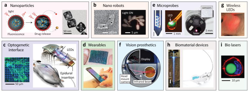
Various implantable photonic devices at the proof-of-concept stage.
a, Nanoparticles for drug delivery and treatment monitoring.
Left, Light-activated drug release. Right, photothermal-polymer-coated gold
nanocages.
b, Nanorobots. Left, Transmission electron microscope image of
an aptamer-gated DNA nanorobot capable of transporting 5-nm gold nanoparticles
(black dots) to cells.
Right, Light-controllable microrobot with nanoscale phototactic
machinery.
c, Optogenetic devices. Left, Optical pattern illuminated on
cultured neuronal cells.
Right, Miniaturized, wireless, soft optoelectronic systems implanted in the
spinal cord for performing optogenetics. d, Wearables. Top, Thermo-chromic
liquid-crystal-based temperature imaging device on the skin. Bottom, Epidermal optoelectronic
device powered by, and communicating with, a smartphone. e, Microprobes. Left,
Multifunctional, implantable optoelectronic device with LEDs and
microelectrodes.
Middle, optoelectronic drug-delivery probe. Right, multifunctional endoscope system based on
transparent bioelectronic sensors and theranostic nanoparticles. f, Vision
prosthetics. Retinal prosthetic system using a head-mounted camera and a goggle,
which projects NIR (880–915 nm) images to photodiode arrays implanted in
the retina.
g, Wirelessly-powered, subcutaneously implanted LED in a
mouse.
h, Biomaterial devices. Left, biodegradable polymer waveguide
implanted in tissue for efficient light delivery. Right, Fibre-pigtailed hydrogel
waveguide implanted in a freely moving mouse for sensing and therapy. i, Human cell
containing an intracellular fluorescent-bead laser (green).
References
-
- Zaret MM, et al. Ocular lesions produced by an optical maser (laser) Science. 1961;134:1525–1526. - PubMed
-
- Goldman L, Wilson RG. Treatment of basal cell epithelioma by laser radiation. JAMA. 1964;189:773–775. - PubMed
-
- Sakimoto T, Rosenblatt MI, Azar DT. Laser eye surgery for refractive errors. Lancet. 2006;367:1432–1447. - PubMed
-
- Marshall J, Trokel S, Rothery S, Krueger R. Long-term healing of the central cornea after photorefractive keratectomy using an exicmer laser. Opthalmology. 1988;95:1411–1421. - PubMed
-
- Solomon KD, et al. LASIK world literature review: quality of life and patient satisfaction. Ophthalmology. 2009;116:691–701. - PubMed
Grants and funding
LinkOut - more resources
Full Text Sources
Other Literature Sources
