. 2017 Apr 25:3:15.
doi: 10.1038/s41531-017-0017-1. eCollection 2017.

Imaging Parkinson's disease below the neck

Affiliations

Imaging Parkinson's disease below the neck

Per Borghammer et al. NPJ Parkinsons Dis. .

Abstract

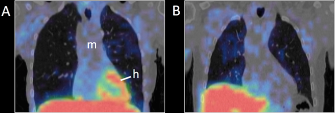
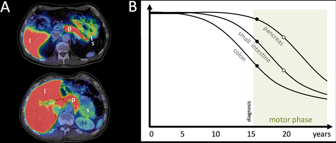
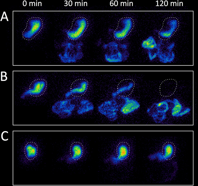
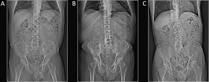
Parkinson's disease is a systemic disorder with widespread and early α-synuclein pathology in the autonomic and enteric nervous systems, which is present throughout the gastrointestinal canal prior to diagnosis. Gastrointestinal and genitourinary autonomic symptoms often predate clinical diagnosis by several years. It has been hypothesized that progressive α-synuclein aggregation is initiated in hyperbranched, non-myelinated neuron terminals, and may subsequently spread via retrograde axonal transport. This would explain why autonomic nerves are so prone to formation of α-synuclein pathology. However, the hypothesis remains unproven and in vivo imaging methods of peripheral organs may be essential to study this important research field. The loss of sympathetic and parasympathetic nerve terminal function in Parkinson's disease has been demonstrated using radiotracers such as 123I-meta-iodobenzylguanidin, 18F-dopamine, and 11C-donepezil. Other radiotracer and radiological imaging methods have shown highly prevalent dysfunction of pharyngeal and esophageal motility, gastric emptying, colonic transit time, and anorectal function. Here, we summarize the methodology and main findings of radio-isotope and radiological modalities for imaging peripheral pathology in Parkinson's disease.


Conflict of interest statement

Professor Brooks have received consultancies from GE Healthcare. Dr Borghammer has received consultancies from F. Hoffmann—La Roche. All other authors declare that they have no competing interests.

Figures

Fig. 1
Fig. 1
123I-MIBG SPECT images superimposed on anatomical CT. a Healthy subject with normal cardiac uptake. b PD patient with severely decreased cardiac uptake. [h heart, m mediastinum]
Fig. 2
Fig. 2
11C-donepezil PET/CT images. a Summed PET images superimposed on anatomical CT in a healthy control (top) and a PD patient (bottom). Note the visually apparent decrease in the small intestine signal. b Hypothetical timeline of parasympathetic denervation during the course of prodromal and manifest PD. Closed and open circles represent 11C-donepezil PET data from de novo (duration 1 year) and moderate stage PD patients (duration 4.5 years), respectively., Shortly after diagnosis, some 11C-donepezil signal loss is seen in the small intestine and colon, but not in the pancreas. Five years after diagnosis, progressive signal loss is seen in the pancreas, small intestine, and colon. [l liver, p pancreas, s small intestine]
Fig. 3
Fig. 3
Gastric emptying scintigraphy images at 0, 30, 60, and 120 min after radioactive meal ingestion. a Healthy control with normal gastric emptying time (T 1/2 72 min). The dashed line designates the stomach. b PD patient with rapid gastric emptying time (T 1/2 26 min). c Vagotomized patient with severely increased gastric emptying time (T 1/2 > 180 min)
Fig. 4
Fig. 4
Radio-opaque marker studies of colonic transit time (CTT) using the 7-day (60 marker) protocol. a Healthy control with rapid CTT (4 markers). b PD patient with increased CTT (28 markers). c PD patient with severely increased CTT (57 markers). Note that the markers are situated mostly in the descending and recto-sigmoid colon suggestive of “outlet obstruction” constipation
Fig. 5
Fig. 5
Thermograms in a healthy control subject (a) and two patients with PD (b). Note the asymmetric hand temperature in the patients. [Figure provided at the courtesy of the Dr Antonio-Rubio and co-authors]

References

    1. Jellinger KA. The pathomechanisms underlying Parkinson’s disease. Expert Rev. Neurother. 2014;14:199–215. doi: 10.1586/14737175.2014.877842. - DOI - PubMed
    1. Beach TG, et al. Multi-organ distribution of phosphorylated alpha-synuclein histopathology in subjects with Lewy body disorders. Acta Neuropathol. 2010;119:689–702. doi: 10.1007/s00401-010-0664-3. - DOI - PMC - PubMed
    1. Gelpi E, et al. Multiple organ involvement by alpha-synuclein pathology in Lewy body disorders. Mov. Disord. 2014;29:1010–1018. doi: 10.1002/mds.25776. - DOI - PubMed
    1. Hilton D, et al. Accumulation of alpha-synuclein in the bowel of patients in the pre-clinical phase of Parkinson’s disease. Acta Neuropathol. 2014;127:235–241. doi: 10.1007/s00401-013-1214-6. - DOI - PubMed
    1. Shannon KM, Keshavarzian A, Dodiya HB, Jakate S, Kordower JH. Is alpha-synuclein in the colon a biomarker for premotor Parkinson’s disease? Evidence from 3 cases. Mov. Disord. 2012;27:716–719. doi: 10.1002/mds.25020. - DOI - PubMed

LinkOut - more resources