. 2016 Dec 19:2:2.
doi: 10.1038/s41526-016-0001-9. eCollection 2016.

Brain structural plasticity with spaceflight

Affiliations

Brain structural plasticity with spaceflight

Vincent Koppelmans et al. NPJ Microgravity. .

Erratum in

Abstract

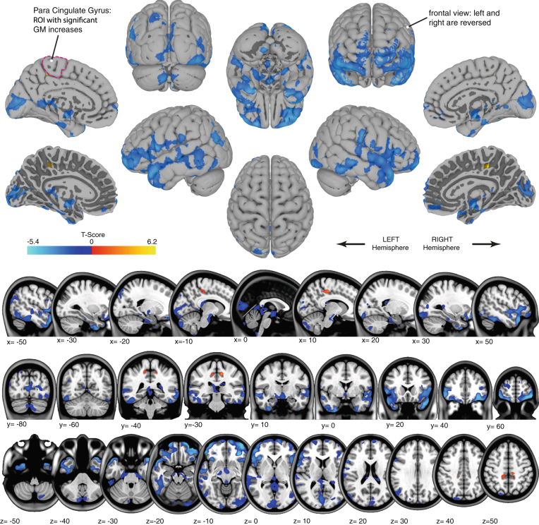
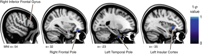
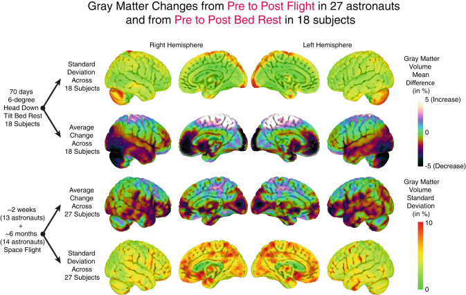
Humans undergo extensive sensorimotor adaptation during spaceflight due to altered vestibular inputs and body unloading. No studies have yet evaluated the effects of spaceflight on human brain structure despite the fact that recently reported optic nerve structural changes are hypothesized to occur due to increased intracranial pressure occurring with microgravity. This is the first report on human brain structural changes with spaceflight. We evaluated retrospective longitudinal T2-weighted MRI scans and balance data from 27 astronauts (thirteen ~2-week shuttle crew members and fourteen ~6-month International Space Station crew members) to determine spaceflight effects on brain structure, and whether any pre to postflight brain changes are associated with balance changes. Data were obtained from the NASA Lifetime Surveillance of Astronaut Health. Brain scans were segmented into gray matter maps and normalized into MNI space using a stepwise approach through subject specific templates. Non-parametric permutation testing was used to analyze pre to postflight volumetric gray matter changes. We found extensive volumetric gray matter decreases, including large areas covering the temporal and frontal poles and around the orbits. This effect was larger in International Space Station versus shuttle crew members in some regions. There were bilateral focal gray matter increases within the medial primary somatosensory and motor cortex; i.e., the cerebral areas where the lower limbs are represented. These intriguing findings are observed in a retrospective data set; future prospective studies should probe the underlying mechanisms and behavioral consequences.


Figures

Fig. 1
Fig. 1
Gray matter changes as a function of spaceflight. Red-to-yellow gradients show regions with significant gray matter volume increase. Blue gradients show regions with significant gray matter decreases. Results are displayed on top of the pial surface of the ICBM MNI brain (surface view; top) or overlaid on the ICBM MNI T1 image (bottom). For the medial surface views, additional surface images that show the border of the white and gray matter are provided (marked with “*”) to show the gray matter changes that are present in deeper sulcal regions. The right side of the image corresponds with the right side of the brain, unless mentioned otherwise. The para cingulate gyrus that shows significant overall gray matter increases from pre- to post-flight is outlined on one of the surface images
Fig. 2
Fig. 2
Focal differences in gray matter volume changes between shuttle astronauts and ISS astronauts. Blue gradients show regions in which ISS astronauts had significantly larger decreases in gray matter than shuttle astronauts from pre to post-flight. Results are overlaid on the MNI152 brain
Fig. 3
Fig. 3
Qualitative comparison of focal gray matter changes with bed rest and spaceflight. Images on the two middle rows show average focal gray matter changes from pre to post-bed rest [18 subjects] and from pre to post-spaceflight [27 subjects] plotted on the ICBM MNI brain. Red-to-yellow gradients indicate gray matter increases whereas blue gradients indicate gray matter decreases. The top and bottom rows show the standard deviation of focal gray matter changes in these samples. This overview is intended for qualitative comparison. No statistical tests were run because of differences in pre-to-post intervals, scan sequence and parameters, and demographic differences

References

    1. Nelson, E. S., Mulugeta, L. & Myers, J. G. Microgravity-induced fluid shift and ophthalmic changes. Life4, 621–665 (2014). PubMed PMID: 25387162. Pubmed Central PMCID: 4284461. - PMC - PubMed
    1. Parihar, V. K., et al. What happens to your brain on the way to Mars. Sci. Adv.1, 1–6 (2015) PubMed PMID: 26180843. Pubmed Central PMCID: 4500198. - PMC - PubMed
    1. Roberts, D. R., et al. Structural brain changes following long-term 6 degrees head-down tilt bed rest as an analog for spaceflight. AJNR Am. J. Neuroradiol.36, 2048–2054 (2015). PubMed PMID: 26185326. - PMC - PubMed
    1. Cohen, H. S., Kimball, K. T., Mulavara, A. P., Bloomberg, J. J. & Paloski, W. H. Posturography and locomotor tests of dynamic balance after long-duration spaceflight. J. Vestib. Res. 22, 191–196 (2012). PubMed PMID: 23142833. - PMC - PubMed
    1. Paloski WH, Bloomberg JJ, Reschke MF, Hamm DL. Spaceflight-induced changes in posture and locomotion. J. Biomech. 1994;27(6):812. doi: 10.1016/0021-9290(94)91366-8. - DOI