

Inflammation of mammary adipose tissue occurs in overweight and obese patients exhibiting early-stage breast cancer
- PMID: 28649659
- PMCID: PMC5460134
- DOI: 10.1038/s41523-017-0015-9
Inflammation of mammary adipose tissue occurs in overweight and obese patients exhibiting early-stage breast cancer
Erratum in
-
Erratum: Inflammation of mammary adipose tissue occurs in overweight and obese patients exhibiting early-stage breast cancer.NPJ Breast Cancer. 2017 Sep 5;3:35. doi: 10.1038/s41523-017-0030-x. eCollection 2017. NPJ Breast Cancer. 2017. PMID: 28884144 Free PMC article.
Abstract
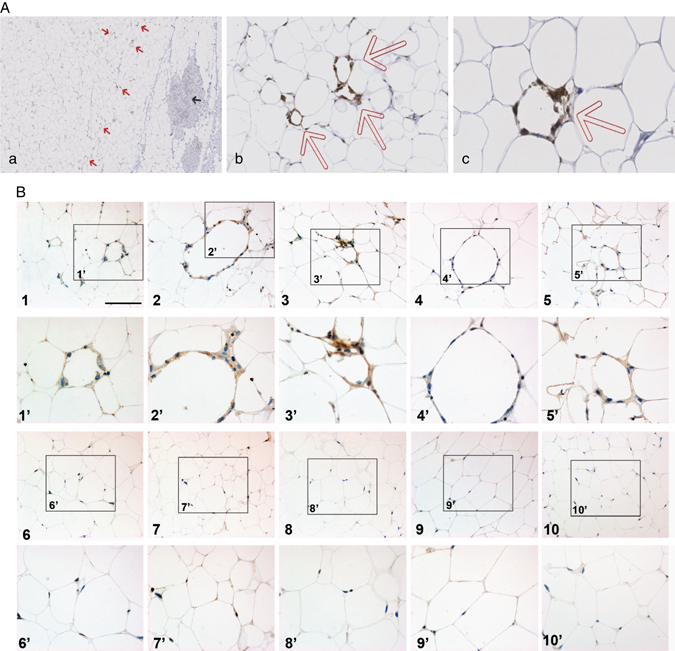
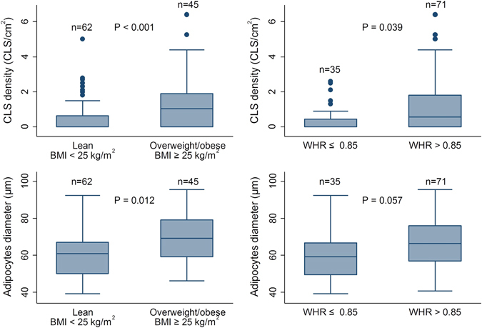
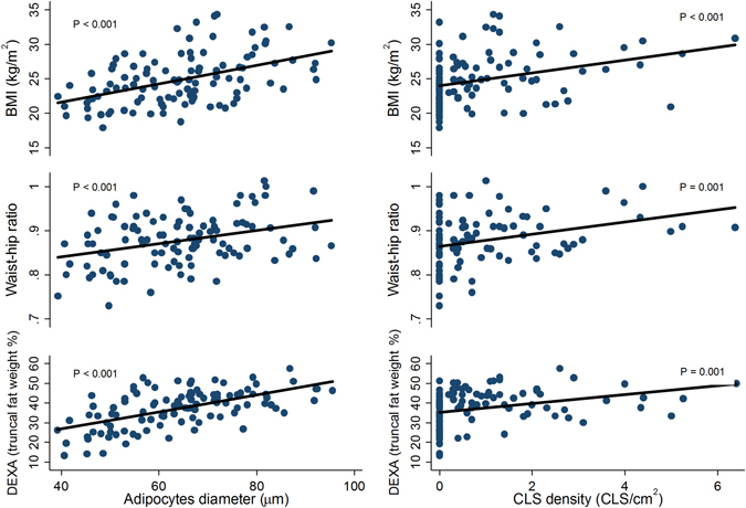
Growing evidence indicates that adiposity is associated with breast cancer risk and negatively affects breast cancer recurrence and survival, a paracrine role of mammary adipose tissue being very likely in this process. In contrast to other adipose depots, occurrence of a sub-inflammatory state of mammary adipose tissue defined by dying adipocytes surrounded by macrophages forming crown-like structures in overweight and obese subjects, remains only partially described. In a general population of breast cancer patients (107 patients) mostly undergoing breast-conserving surgery, we found a positive association between patient's body composition, breast adipocytes size, and presence of crown-like structures in mammary adipose tissue close to the tumor. Overweight (BMI: 25.0-29.9 kg/m2) and obese (BMI ≥ 30.0 kg/m2) patients have 3.2 and 6.9 times higher odds ratio of crown-like structures respectively, compared with normal weight patients. The relatively small increase in adipocyte size in crown-like structures positive vs. negative patients suggests that mammary adipose tissue inflammation might occur early during hypertrophy. Our results further highlight that body mass index is an adequate predictor of the presence of crown-like structures in mammary adipose tissue among postmenopausal women, whereas in premenopausal women truncal fat percentage might be more predictive, suggesting that mammary adipose tissue inflammation is more likely to occur in patients exhibiting visceral obesity. Finally, the presence of crown-like structures was positively associated with systemic markers such as the Triglyceride/High-density lipoprotein-cholesterol ratio serum C-reactive protein and glucose/(HbA1c) glycated Haemoglobin. These compelling results demonstrate that excess adiposity, even in overweight patients, is associated with mammary adipose tissue inflammation, an event that could contribute to breast cancer development and progression.
Conflict of interest statement
The authors declare that they have no competing interests.
Figures

References
LinkOut - more resources
Full Text Sources
Other Literature Sources
Research Materials
