. 2017 Aug 1;127(8):2904-2915.
doi: 10.1172/JCI75005. Epub 2017 Jun 26.

Inflammatory Ly6Chi monocytes and their conversion to M2 macrophages drive atherosclerosis regression

Affiliations

Inflammatory Ly6Chi monocytes and their conversion to M2 macrophages drive atherosclerosis regression

Karishma Rahman et al. J Clin Invest. .

Abstract

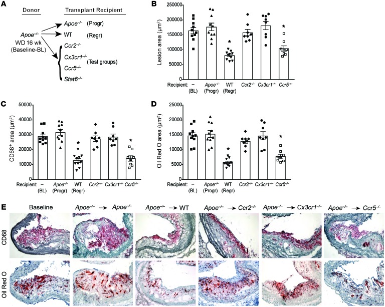
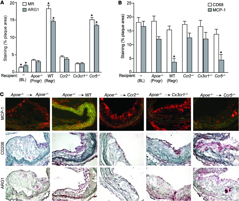
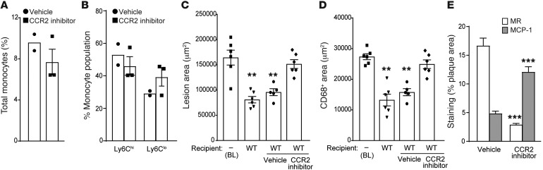
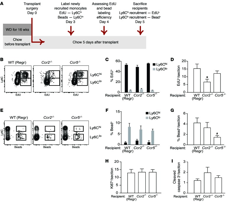
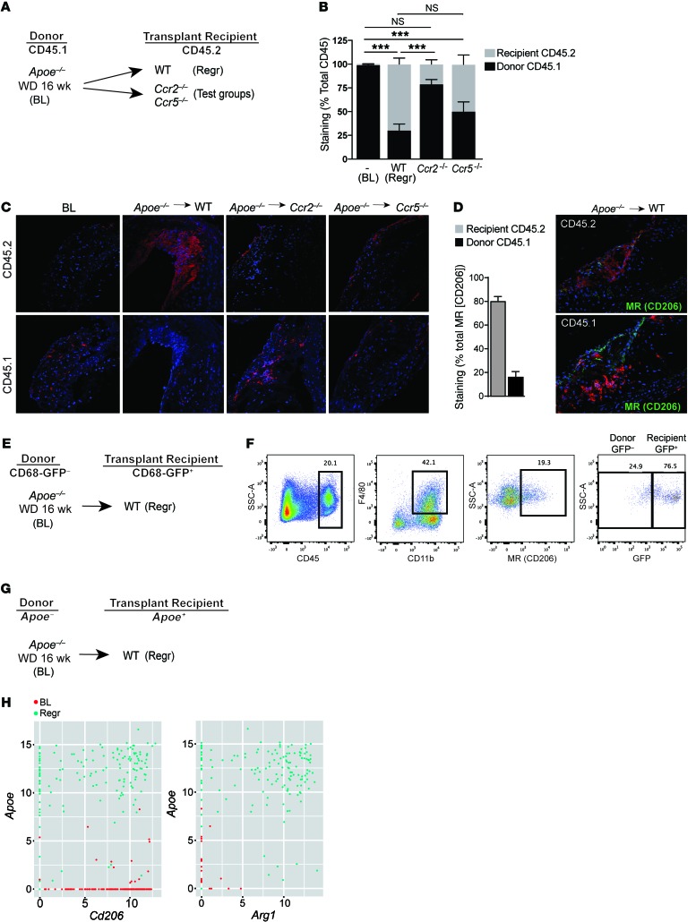
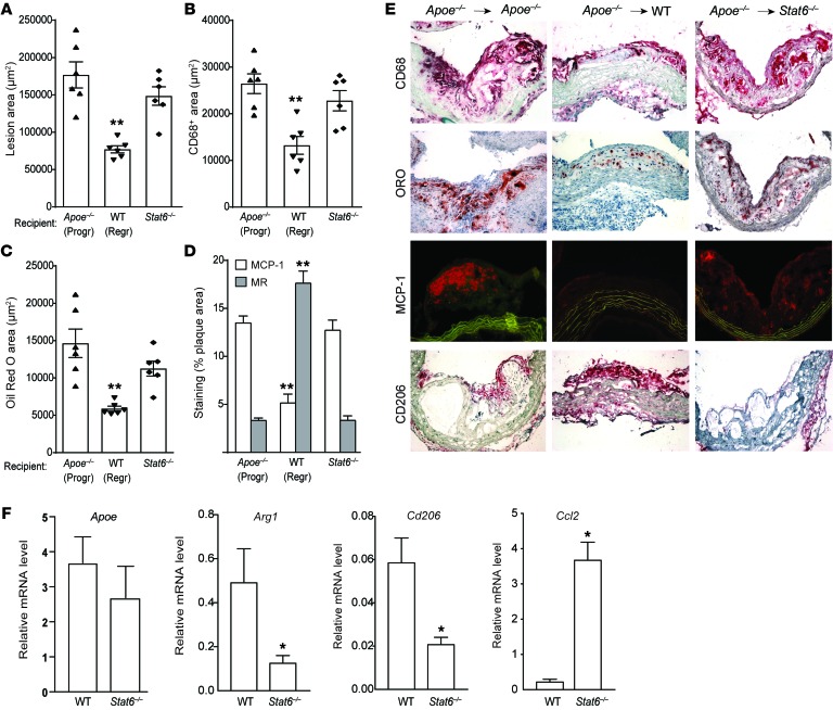
Atherosclerosis is a chronic inflammatory disease, and developing therapies to promote its regression is an important clinical goal. We previously established that atherosclerosis regression is characterized by an overall decrease in plaque macrophages and enrichment in markers of alternatively activated M2 macrophages. We have now investigated the origin and functional requirement for M2 macrophages in regression in normolipidemic mice that received transplants of atherosclerotic aortic segments. We compared plaque regression in WT normolipidemic recipients and those deficient in chemokine receptors necessary to recruit inflammatory Ly6Chi (Ccr2-/- or Cx3cr1-/-) or patrolling Ly6Clo (Ccr5-/-) monocytes. Atherosclerotic plaques transplanted into WT or Ccr5-/- recipients showed reduced macrophage content and increased M2 markers consistent with plaque regression, whereas plaques transplanted into Ccr2-/- or Cx3cr1-/- recipients lacked this regression signature. The requirement of recipient Ly6Chi monocyte recruitment was confirmed in cell trafficking studies. Fate-mapping and single-cell RNA sequencing studies also showed that M2-like macrophages were derived from newly recruited monocytes. Furthermore, we used recipient mice deficient in STAT6 to demonstrate a requirement for this critical component of M2 polarization in atherosclerosis regression. Collectively, these results suggest that continued recruitment of Ly6Chi inflammatory monocytes and their STAT6-dependent polarization to the M2 state are required for resolution of atherosclerotic inflammation and plaque regression.


Conflict of interest statement

Conflict of interest: V. Gusarova and J. Gromada are employees and shareholders of Regeneron Pharmaceuticals. N.M. Girgis has been an employee of Janssen Research and Development and is currently an employee of Constellation Pharmaceuticals.

Figures

Figure 1
Figure 1. CCR2 and CX3CR1 are required for plaque regression.
Analysis of aortic arch plaques from Apoe–/– mice on 16-week WD (baseline [BL]; n = 10) or 5 days after transplantation into Apoe–/– (progression [Progr]; n = 10), WT (regression [Regr]; n = 10), or chemokine receptor–KO recipient mice (Ccr2–/–, Cx3cr1–/–, or Ccr5–/–; n = 8). (A) Schematic of transplant experiments. Quantification of (B) lesion area, (C) immunohistochemical staining for the macrophage marker CD68, and (D) Oil Red O staining of neutral lipids. *P < 0.001 compared with BL and Apoe–/– progression groups using 1-way ANOVA with Dunnett’s multiple comparisons testing. (E) Representative sections of aortic transplant segments from BL and the recipient groups stained with anti-CD68 antibody and Oil Red O, imaged at ×40 magnification.
Figure 2
Figure 2. CCR2 and CX3CR1 are required for enrichment of M2 macrophages in the regressing plaque.
Quantification of immunohistochemical staining for (A) markers of M2 macrophages MR and ARG1 and (B) the macrophage marker CD68 and an M1 macrophage marker MCP-1, in aortic arch plaques from Apoe–/– mice on a 16-week WD (BL; n = 10) or 5 days after transplantation into Apoe–/– (Progr; n = 10), WT (Regr; n = 10), or chemokine receptor–deficient recipient mice (Ccr2–/–, Cx3cr1–/– or Ccr5–/–; n = 8). *P < 0.01 compared with Apoe–/– progression group using 1-way ANOVA with Dunnett’s multiple comparisons testing. (C) Representative images of aortic plaques stained for MCP-1, CD206 (MR), and ARG1, imaged at ×40 magnification.
Figure 3
Figure 3. Effect of CCR2 inhibition on atherosclerosis regression and M2 macrophage enrichment.
Effect of CCR2 inhibitor on the frequency of (A) total monocytes and (B) Ly6Chi and Ly6Clo monocyte subsets in the blood of recipient mice (vehicle, n = 2, and CCR2 inhibitor, n = 3). (CE) Quantification of (C) lesion area, (D) the macrophage marker CD68, and (E) MR and MCP-1 staining in aortic arch plaques from Apoe–/– mice on 16-week WD (BL; n = 6) or 3 days after transplant into WT mice (regression environment) that were untreated (n = 6) or treated with vehicle (n = 5) or a CCR2 inhibitor (n = 6). For C and D: **P < 0.001 compared with BL using 1-way ANOVA with Dunnett’s multiple comparisons testing. For E, ***P < 0.0001, CCR2 inhibitor group compared with vehicle group using unpaired t testing.
Figure 4
Figure 4. Macrophage dynamics show Ly6chi monocyte recruitment is the key kinetic change impairing regression in Ccr2–/– recipient mice.
Aortic arches from Apoe–/– donors fed WD for 14 weeks were transplanted into recipients. (A) Schematic of timeline for EdU and bead injections into recipient mice to assess recruitment of Ly6Chi and Ly6Clo monocytes, respectively, into transplanted aortic arches under regression conditions. (B) Representative flow cytometry plots of CD45+CD115+ circulating monocytes showing Ly6C versus EdU, and (C) quantification of EdU incorporation in circulating Ly6Chi versus Ly6Clo populations showing that EdU is preferentially incorporated into Ly6Chi monocytes in WT, Ccr2–/–, and Ccr5–/– mice (n = 4–8 per group). (D) Analysis of EdU+ cells/section of atherosclerotic plaques transplanted into WT, Ccr2–/–, or Ccr5–/– recipients showing significantly reduced recruitment into plaques of Ly6ChiEdU+ cells into Ccr2–/– compared with WT recipients. (E) Representative flow cytometry plots of CD45+CD115+ circulating monocytes showing Ly6C versus beads, and (F) quantification of bead incorporation in circulating Ly6Chi versus Ly6Clo monocyte populations showing that beads are preferentially incorporated into Ly6Clo monocytes in WT, Ccr2–/–, and Ccr5–/– mice (n = 8–10 per group). (G) Analysis of bead+ cells/section of atherosclerotic plaques transplanted into WT, Ccr2–/–, or Ccr5–/– recipients showing significantly reduced recruitment of Ly6Clo bead+ cells into Ccr5–/– compared with WT mice. Quantification in plaque sections of (H) Ki67 (to assess proliferation) and (I) cleaved caspase 3 (to assess apoptosis) showed no significant differences in proliferation or apoptosis between transplant recipient groups. Quantifications in D, G, H, and I were done in aortic arch plaques from mice 5 days after transplantation (n = 8–9 per group). #P = 0.05, *P < 0.05 when compared with WT group using 1-way ANOVA with Dunnett’s multiple comparisons testing.
Figure 5
Figure 5. CCR2 deficiency in recipient leukocytes impairs their recruitment to atherosclerotic plaques, where they normally display M2 characteristics.
(A) Schematic of CD45.1 (donor) to CD45.2 (recipient) aortic transplantation experiments with (B) quantification of immunohistochemical staining of CD45.1 and CD45.2 in aortic arch plaques from Apoe–/– mice on 14-week WD (BL; n = 7) or 5 days after transplant into WT mice (Regr; n = 8), or chemokine receptor–KO recipient mice (Ccr2–/– or Ccr5–/–; n = 8); ***P < 0.001 for the indicated comparisons group using 1-way ANOVA with Dunnett’s multiple comparisons testing. (C) Representative images of aortic plaques stained for CD45.1 and CD45.2, imaged at ×40 quantification. (D) Quantification of immunohistochemical staining of MR+ with CD45.2 or CD45.1 showing 80.72% ± 3.597% MR+ cells originate from recipient CD45.2 mice (n = 3) with representative images at ×40 magnification. (E) Schematic of transplantation experiment using CD68-GFP reporter mice to mark recipient monocytes/macrophages. (F) Representative flow cytometry plots showing that a majority of CD45+CD11b+F4/80+MR+ macrophages are GFP+ in aortic arches harvested 5 days after transplantation (n = 3). (G) Schematic of transplant experiment for single cell RNA-seq experiments, with (H) scatter plots showing, at a single-cell level, that cells expressing high levels of Apoe (i.e., recipient) are also positive for high levels of M2 macrophage markers Cd206 (MR) and Arg1 in the population of Cd11b+F4/80+ macrophages isolated from aortic arches 3 days after transplantation.
Figure 6
Figure 6. Lack of STAT6 in recipient mice prevents M2 macrophage enrichment of plaques and impairs atherosclerosis regression.
Analysis of aortic arch plaques from mice 3 days after transplantation into Apoe–/– (Progr; n = 6), WT (Regr; n = 6), or Stat6–/– (n = 6) mice for (A) lesion area, (B) immunohistochemical staining for the macrophage marker CD68, (C) Oil red O staining for neutral lipid, and (D) immunohistochemical staining for the M1 macrophage marker MCP-1 and the M2 macrophage marker MR (CD206); **P < 0.001 compared with Apoe–/– progression group using 1-way ANOVA with Dunnett’s multiple comparisons testing. (E) Representative images of aortic plaques stained for CD68, Oil red O (ORO), MCP-1, and CD206, imaged at ×40 magnification. (F) qRT-PCR analysis of mRNA expression of newly recruited monocyte-derived macrophage marker (Apoe), CD206 (M2) (Arg1 and Cd206), and M1 (Ccl2) macrophage markers in CD68+ cells laser captured from the aortic arch plaques 3 days after transplant into WT and Stat6–/– recipient mice (n = 4–7 per group). *P < 0.05 using unpaired t testing.

Similar articles

Cited by

References

    1. Williams KJ, Feig JE, Fisher EA. Rapid regression of atherosclerosis: insights from the clinical and experimental literature. Nat Clin Pract Cardiovasc Med. 2008;5(2):91–102. doi: 10.1038/ncpcardio1086. - DOI - PubMed
    1. Feig JE, et al. Reversal of hyperlipidemia with a genetic switch favorably affects the content and inflammatory state of macrophages in atherosclerotic plaques. Circulation. 2011;123(9):989–998. doi: 10.1161/CIRCULATIONAHA.110.984146. - DOI - PMC - PubMed
    1. Hewing B, Parathath S, Mai CK, Fiel MI, Guo L, Fisher EA. Rapid regression of atherosclerosis with MTP inhibitor treatment. Atherosclerosis. 2013;227(1):125–129. doi: 10.1016/j.atherosclerosis.2012.12.026. - DOI - PMC - PubMed
    1. Rayner KJ, et al. Antagonism of miR-33 in mice promotes reverse cholesterol transport and regression of atherosclerosis. J Clin Invest. 2011;121(7):2921–2931. doi: 10.1172/JCI57275. - DOI - PMC - PubMed
    1. Feig JE, et al. Regression of atherosclerosis is characterized by broad changes in the plaque macrophage transcriptome. PLoS One. 2012;7(6):e39790. doi: 10.1371/journal.pone.0039790. - DOI - PMC - PubMed

MeSH terms