. 2017 Aug 1;127(8):2968-2981.
doi: 10.1172/JCI93868. Epub 2017 Jun 26.

Preexisting endothelial cells mediate cardiac neovascularization after injury

Affiliations

Preexisting endothelial cells mediate cardiac neovascularization after injury

Lingjuan He et al. J Clin Invest. .

Abstract

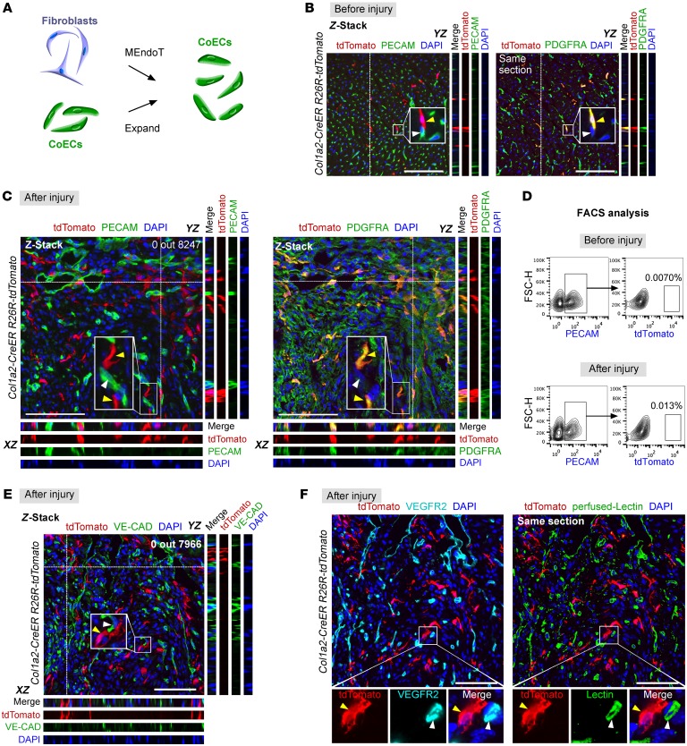
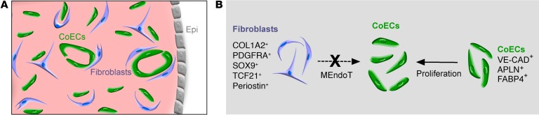
The mechanisms that promote the generation of new coronary vasculature during cardiac homeostasis and after injury remain a fundamental and clinically important area of study in the cardiovascular field. Recently, it was reported that mesenchymal-to-endothelial transition (MEndoT) contributes to substantial numbers of coronary endothelial cells after myocardial infarction. Therefore, the MEndoT has been proposed as a paradigm mediating neovascularization and is considered a promising therapeutic target in cardiac regeneration. Here, we show that preexisting endothelial cells mainly beget new coronary vessels in the adult mouse heart, with essentially no contribution from other cell sources through cell-lineage transdifferentiation. Genetic-lineage tracing revealed that cardiac fibroblasts expand substantially after injury, but do not contribute to the formation of new coronary blood vessels, indicating no contribution of MEndoT to neovascularization. Moreover, genetic-lineage tracing with a pulse-chase labeling strategy also showed that essentially all new coronary vessels in the injured heart are derived from preexisting endothelial cells, but not from other cell lineages. These data indicate that therapeutic strategies for inducing neovascularization should not be based on targeting presumptive lineage transdifferentiation such as MEndoT. Instead, preexisting endothelial cells appear more likely to be the therapeutic target for promoting neovascularization and driving heart regeneration after injury.


Conflict of interest statement

Conflict of interest: The authors have declared that no conflict of interest exists.

Figures

Figure 1
Figure 1. COL1A2+ fibroblasts do not adopt the endothelial cell fate after injury.
(A) Models explaining new vessel sources: MEndoT and self-expansion. CoECs, coronary endothelial cells. (B) Z-stack confocal images of heart sections stained for tdTomato, PECAM, and PDGFRA. Col1a2-CreER R26R-tdTomato mice were treated with tamoxifen 2 weeks before analysis. YZ indicates signals from dotted lines on Z-stack images. Yellow arrowheads indicate PDGFRA+tdTomato+ fibroblasts; white arrowheads indicate PECAM+tdTomato endothelial cells. (C) Immunostaining for tdTomato, PDGFRA, and PECAM on sections of postinjury hearts. tdTomato+ cells express fibroblast marker PDGFRA+ (yellow arrowheads), but not endothelial cell marker PECAM (white arrowheads). (D) Flow cytometric analysis of percentage of PECAM+ endothelial cells labeled by Col1a2-CreER transgene (tdTomato+). (E) Immunostaining for VE-CAD and tdTomato on sections of injured heart. tdTomato+ cells (yellow arrowheads) are close to, but were not identified as VE-CAD+ endothelial cells (white arrowheads) in the injured heart. (F) Immunostaining for VEGFR2 and tdTomato on heart sections perfused with FITC-labeled BS1 lectin. tdTomato+ cells (yellow arrowheads) are not VEGFR2+lectin+ vascular endothelial cells (white arrowheads). Scale bars: 100 μm. Each image is representative of 4 individual hearts.
Figure 2
Figure 2. COL1A2+ fibroblasts do not contribute to coronary endothelial cells after cardiac injury.
(A) Schematic figure showing strategy for generation of Col1a2-2A-CreER allele. (B and C) Whole-mount fluorescence images showing tdTomato in hearts before or after injury. (D and E) Immunostaining for tdTomato, PDGFRA, and PECAM (D) or VE-CAD (E) on sections of injured heart. Boxed regions are magnified in bottom panels. Arrowheads point to tdTomato+PDGFRA+PECAM (D) or tdTomato+PDGFRA+VE-CAD cells (E). XZ indicates signals from dotted line in Z-stack images. (F) Quantification of the percentage of tdTomato+ cells in different lineages (PDGFRA+, PECAM+, or VE-CAD+ cell populations). Data are represented as mean ± SEM. n = 4. (G) Immunostaining for tdTomato and DDR2 on heart sections before or after injury. (H) Flow cytometric analysis of the percentage of tdTomato+ endothelial cells. FSC-H, forward scatter–height. Scale bars: 200 μm (B, C); 100 μm (D, E, G).
Figure 3
Figure 3. PDGFRA+ fibroblasts do not contribute to coronary endothelial cells after cardiac injury.
(A) Schematic figure showing strategy for generation of Pdgfra-DreER allele. (B) Schematic figure showing genetic-lineage–tracing strategy for PDGFRA+ cells by Dre-rox recombination. (C and D) Flow cytometric analysis of the percentage of tdTomato+ fibroblasts (C) or tdTomato+ endothelial cells (D). (E) Whole-mount fluorescence image showing tdTomato in hearts before and after injury. (F–J) Immunostaining for tdTomato, PDGFRA, PECAM, or VE-CAD on sections of injured heart. Boxed regions in F are magnified in GJ. Arrowheads point to tdTomato+PDGFRA+PECAM (G, H) or tdTomato+PDGFRA+VE-CAD cells (I and J). YZ indicates signals from dotted line in Z-stack images in GJ. (K) Quantification of the percentage of tdTomato+ cells in different lineages (PECAM+, VE-CAD+, or PDGFRA+ cell populations). Data are represented as mean ± SEM. n = 4. Scale bars: 200 μm (E); 100 μm in (FJ).
Figure 4
Figure 4. Cardiac fibroblasts expand after injury without giving rise to endothelial cells.
(A and C) Whole-mount fluorescence view of Sox9-CreER R26R-tdTomato hearts before (A) and after injury (C). Inserts indicate bright-field view of hearts. (B and D) Immunostaining for tdTomato, PDGFRA, and PECAM on heart sections before (B) and after injury (D). XZ and YZ indicate signals from dotted lines on Z-stack images. Yellow arrowheads indicate PDGFRA+tdTomato+ fibroblasts; white arrowheads indicate PECAM+tdTomato endothelial cells. (E) Flow cytometric analysis of percentage of tdTomato+ cells in PDGFRA+ cell population. (F) Flow cytometric analysis of tdTomato+ cells in PECAM+ endothelial cells from heart before or after injury. (G) Immunostaining for tdTomato and EdU or Ki67 on heart sections before or 3 days after injury. Arrowheads indicate proliferating tdTomato+ cells. (H) Quantification of percentage of proliferating tdTomato+ cells. Data are represented as mean ± SEM. n = 4. *P < 0.05, 2-tailed Student’s t test. Scale bars: 1 mm (A, C); 100 μm (B, D, G).
Figure 5
Figure 5. Establishment of pulse-chase strategy for labeling of coronary endothelial cells.
(A) Schematic figure showing the pulse-chase experimental strategy for measuring refreshment of nonvascular cells. (B) Experimental design for tamoxifen induction, myocardial IR, and tissue analysis. wk, postnatal weeks. (C) Immunostaining for tdTomato and PDGFRA on tissue sections shows that these Cre lines do not label fibroblasts before injury. Each image is representative of 4 individual samples. Scale bars: 100 μm.
Figure 6
Figure 6. Coronary vessels in the injured heart are derived from preexisting coronary vessels.
(A, C, E, G) Immunostaining for tdTomato and PECAM or VE-CAD on heart sections before and after injury. Arrowheads indicate labeled endothelial cells (tdTomato+PECAM+ or tdTomato+VE-CAD+). (B, D, F, H) Quantification of the percentage of tdTomato+ endothelial cells (ECs) in PECAM+ or VE-CAD+ endothelial cells in hearts before injury and 7 or 14 days after injury. Data are represented as mean ± SEM. n = 4. ANOVA test was used. Scale bars: 50 μm.
Figure 7
Figure 7. Preexisting coronary endothelial cells but not fibroblasts contribute to blood vessels after injury.
(A) Image showing coronary endothelial cells (CoECs) and fibroblasts in the adult heart. Epi, epicardium. (B) Image showing contribution of different lineages to coronary endothelial cells after injury.

Comment in

  • The relationship between cardiac endothelium and fibroblasts: it’s complicated

References

    1. Hansson GK. Inflammation, atherosclerosis, and coronary artery disease. N Engl J Med. 2005;352(16):1685–1695. doi: 10.1056/NEJMra043430. - DOI - PubMed
    1. Lin YD, et al. Instructive nanofiber scaffolds with VEGF create a microenvironment for arteriogenesis and cardiac repair. Sci Transl Med. 2012;4(146):146ra109. - PubMed
    1. Riley P. Developmental biology: Plumbing the heart. Nature. 2010;464(7288):498–499. doi: 10.1038/464498a. - DOI - PubMed
    1. Kurian L, et al. Conversion of human fibroblasts to angioblast-like progenitor cells. Nat Methods. 2013;10(1):77–83. - PMC - PubMed
    1. Moore-Morris T, et al. Resident fibroblast lineages mediate pressure overload-induced cardiac fibrosis. J Clin Invest. 2014;124(7):2921–2934. doi: 10.1172/JCI74783. - DOI - PMC - PubMed