

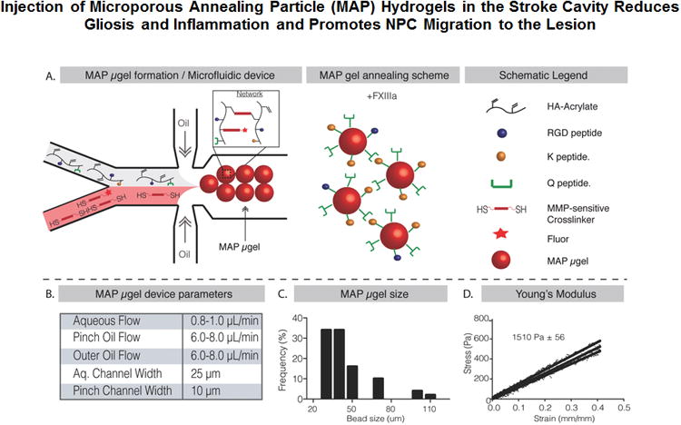
Injection of Microporous Annealing Particle (MAP) Hydrogels in the Stroke Cavity Reduces Gliosis and Inflammation and Promotes NPC Migration to the Lesion
- PMID: 28650574
- PMCID: PMC5595584
- DOI: 10.1002/adma.201606471
Injection of Microporous Annealing Particle (MAP) Hydrogels in the Stroke Cavity Reduces Gliosis and Inflammation and Promotes NPC Migration to the Lesion
Abstract
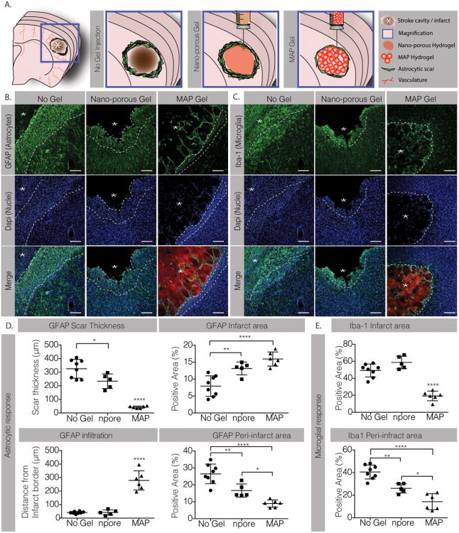
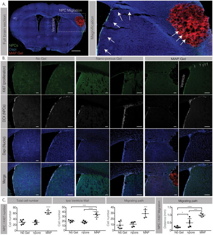
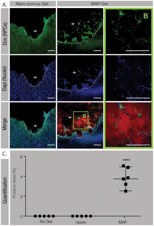
With the number of deaths due to stroke decreasing, more individuals are forced to live with crippling disability resulting from the stroke. To date, no therapeutics exist after the first 4.5 h after the stroke onset, aside from rest and physical therapy. Following stroke, a large influx of astrocytes and microglia releasing proinflammatory cytokines leads to dramatic inflammation and glial scar formation, affecting brain tissue's ability to repair itself. Pathological conditions, such as a stroke, trigger neural progenitor cells (NPCs) proliferation and migration toward the damaged site. However, these progenitors are often found far from the cavity or the peri-infarct tissue. Poststroke tissue remodeling results in a compartmentalized cavity that can directly accept a therapeutic material injection. Here, this paper shows that the injection of a porous hyaluronic acid hydrogel into the stroke cavity significantly reduces the inflammatory response following stroke while increasing peri-infarct vascularization compared to nonporous hydrogel controls and stroke only controls. In addition, it is shown that the injection of this material impacts NPCs proliferation and migration at the subventricular zone niche and results, for the first time, in NPC migration into the stroke site.
Keywords: inflammation; neural progenitor cells; particle hydrogels; porous hydrogels; stroke.
© 2017 WILEY-VCH Verlag GmbH & Co. KGaA, Weinheim.
Figures
References
-
- Go AS, Mozaffarian D, Roger VL, Benjamin EJ, Berry JD, Blaha MJ, Dai S, Ford ES, Fox CS, Franco S, Fullerton HJ, Gillespie C, Hailpern SM, Heit JA, Howard VJ, Huffman MD, Judd SE, Kissela BM, Kittner SJ, Lackland DT, Lichtman JH, Lisabeth LD, Mackey RH, Magid DJ, Marcus GM, Marelli A, Matchar DB, McGuire DK, Mohler ER, 3rd, Moy CS, Mussolino ME, Neumar RW, Nichol G, Pandey DK, Paynter NP, Reeves MJ, Sorlie PD, Stein J, Towfighi A, Turan TN, Virani SS, Wong ND, Woo D, Turner MB C American Heart Association Statistics, S Stroke Statistics. Circulation. 2014;129:e28. - PMC - PubMed
-
- Donnan GA, Fisher M, Macleod M, Davis SM. Lancet. 2008;371:1612. - PubMed
-
- Kim FG, Smith JT, Reeves EE, Navalkele MJ, Grotta DD, Grau-Sepulveda JC, Hernandez MV, Peterson AF, Schwamm ED, Saver JL. Circulation. 2016
MeSH terms
Substances
Grants and funding
LinkOut - more resources
Full Text Sources
Other Literature Sources
