. 2017 Jun 26;12(6):e0179759.
doi: 10.1371/journal.pone.0179759. eCollection 2017.

Accelerated oxygen-induced retinopathy is a reliable model of ischemia-induced retinal neovascularization

Affiliations

Accelerated oxygen-induced retinopathy is a reliable model of ischemia-induced retinal neovascularization

Pilar Villacampa et al. PLoS One. .

Abstract

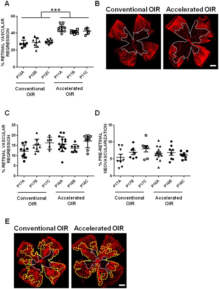
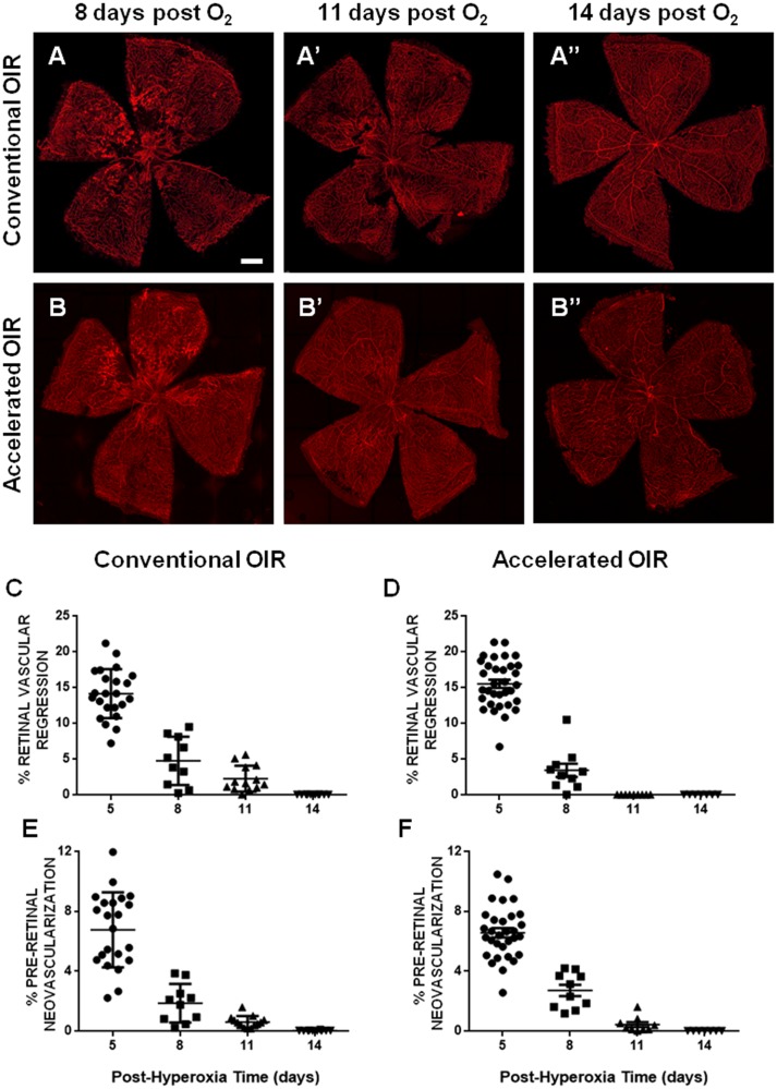
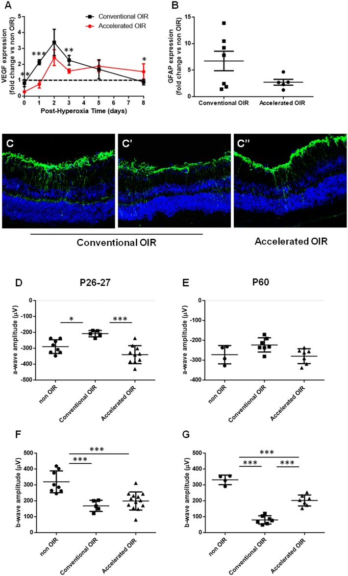
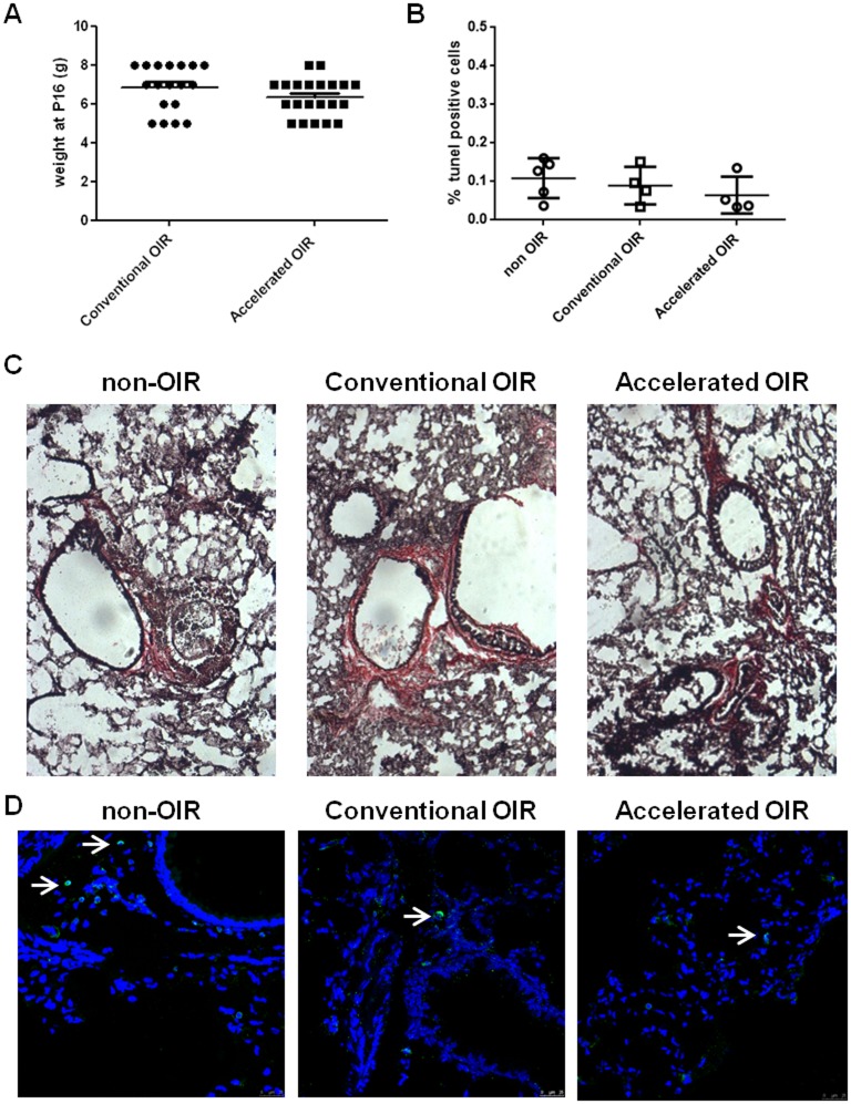
Retinal ischemia and pathological angiogenesis cause severe impairment of sight. Oxygen-induced retinopathy (OIR) in young mice is widely used as a model to investigate the underlying pathological mechanisms and develop therapeutic interventions. We compared directly the conventional OIR model (exposure to 75% O2 from postnatal day (P) 7 to P12) with an alternative, accelerated version (85% O2 from P8 to P11). We found that accelerated OIR induces similar pre-retinal neovascularization but greater retinal vascular regression that recovers more rapidly. The extent of retinal gliosis is similar but neuroretinal function, as measured by electroretinography, is better maintained in the accelerated model. We found no systemic or maternal morbidity in either model. Accelerated OIR offers a safe, reliable and more rapid alternative model in which pre-retinal neovascularization is similar but retinal vascular regression is greater.


Conflict of interest statement

Competing Interests: I have read the journal's policy and the authors of this manuscript have the following competing interests: U.F. Luhmann is an employee of F. Hoffmann-La Roche Ltd. The other authors report no conflicts. There are no patents, products in development or marketed products to declare. This does not alter our adherence to all the PLOS ONE policies on sharing data and materials, as detailed online in the guide for authors.

Figures

Fig 1
Fig 1. Retinal vasculature regression and pre-retinal neovascularization following hyperoxia and 5 days in room air.
(A) The extent of oxygen-induced retinal vascular regression was consistently greater (in 3 independent experimental groups) in the accelerated protocol (85% O2 from P8 to P11) than in the conventional protocol (75% O2 from P7 to P12). No significant differences were evident between independent experimental groups within each protocol, and the variances were similar (Barlett's test, p = 0.6429 for retinal vascular regression and p = 0.1415 for pre-retinal neovascularization). (B) Representative images show the extent of retinal vascular regression (delineated in white) in isolectin B4-stained flat-mounted retinas of mice in the conventional and the accelerated OIR protocols. Five days following return to room air, the extents of both persistent retinal vascular regression (C) and pre-retinal neovascularization (D) were similar in retinas of mice in both protocols. (E) Representative images of flat-mounted retinas of mice from the conventional and accelerated protocols illustrate the area of persistent retinal vascular regression (delineated in white), and the area of pre-retinal neovascularization (delineated in yellow). Scale bars: 0.5 mm. n = 6–14 per group. Data are expressed as means ± SEM.
Fig 2
Fig 2. Time courses of retinal vasculature regeneration and pre-retinal neovascular regression.
(A, B) Representative flat-mounted retinas from mice exposed to conventional OIR (75% O2, upper panel) or accelerated OIR (85% O2, lower panel) at 8 (A, B), 11 (A’, B’) and 14 days after the end of hyperoxia (A”,B”), showing progressive retinal vascular regeneration, and regression of the neovascular tufts. (C, D) Analysis of the persistent retinal vascular regression demonstrated more rapid vascular regeneration in the accelerated protocol at 11 days post-hyperoxia (p = 0.015). (E, F) No differences were found in the kinetics of regression of neovascular lesions between the two experimental conditions. Scale bars: 0.4 mm. n = 7–13 per group. Data are expressed as means ± SEM.
Fig 3
Fig 3. Molecular features and neuroretinal function.
(A) Retinal VEGF was measured by qPCR at intervals (0,1,2,3,5 and 8 days) following exposure to hyperoxia (n = 4–7 per group. (B) GFAP expression measured by qPCR (n = 5–7 per group). (C) Representative images of GFAP immunostaining in retinal sections from mice after the conventional and the accelerated protocol. (C) and (C’) show the variability within the conventional OIR group. (D to G) Electroretinographic (ERG) a-wave and b-wave amplitudes in scotopic conditions were measured at P26-27 (D, E) and at P60 (F, G) in mice previously exposed to the conventional and accelerated OIR protocols, and in age-matched non-OIR controls (n = 4–13 per group). Data are expressed as means ± SEM.
Fig 4
Fig 4. Histological analysis of lungs from nursing mothers after exposure to hyperoxia.
(A) Body weight of pups at P16. (B) Alveolar cell apoptosis indicated by TUNEL positive cells in lung cryosections from non-OIR control mice and mice after conventional or accelerated OIR. (C) Van Gieson staining for collagen in lung cryosections shows no fibrotic lesions in any of the groups analyzed. Original magnification: 20x. (D) CD45-staining of cryosections demonstrates few CD45-positive cells in the lungs of non-OIR control animals and those after conventional or accelerated OIR.

References

    1. Smith LE, Wesolowski E, McLellan A, Kostyk SK, D'Amato R, Sullivan R, et al. Oxygen-induced retinopathy in the mouse. Invest Ophthalmol Vis Sci. 1994;35:101–11. - PubMed
    1. Connor KM, Krah NM, Dennison RJ, Aderman CM, Chen J, Guerin KI, et al. Quantification of oxygen-induced retinopathy in the mouse: a model of vessel loss, vessel regrowth and pathological angiogenesis. Nat Protoc. 2009;4:1565–73. doi: 10.1038/nprot.2009.187 - DOI - PMC - PubMed
    1. Kubota Y, Takubo K, Shimizu T, Ohno H, Kishi K, Shibuya M, et al. M-CSF inhibition selectively targets pathological angiogenesis and lymphangiogenesis. The Journal of experimental medicine. 2009;206:1089–102. doi: 10.1084/jem.20081605 - DOI - PMC - PubMed
    1. Okuno Y, Nakamura-Ishizu A, Otsu K, Suda T, Kubota Y. Pathological neoangiogenesis depends on oxidative stress regulation by ATM. Nat Med. 2012;18:1208–16. doi: 10.1038/nm.2846 - DOI - PubMed
    1. Schneider CA, Rasband WS, Eliceiri KW. NIH Image to ImageJ: 25 years of image analysis. Nature Methods. 2012;9(7):671–5. - PMC - PubMed

MeSH terms

Substances