. 2017 Jun 26;13(6):e1006451.
doi: 10.1371/journal.ppat.1006451. eCollection 2017 Jun.

A human endogenous retrovirus-derived gene that can contribute to oncogenesis by activating the ERK pathway and inducing migration and invasion

Affiliations

A human endogenous retrovirus-derived gene that can contribute to oncogenesis by activating the ERK pathway and inducing migration and invasion

Cécile Lemaître et al. PLoS Pathog. .

Abstract

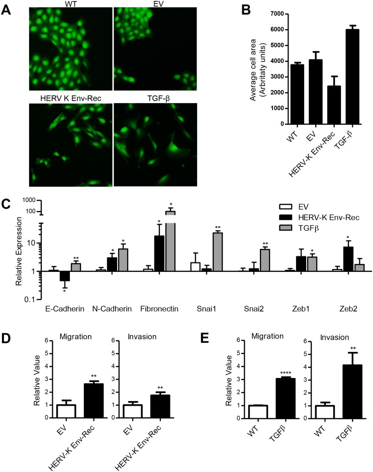
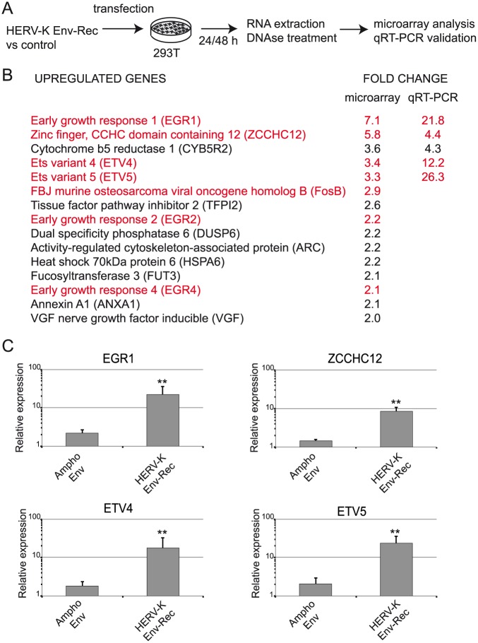
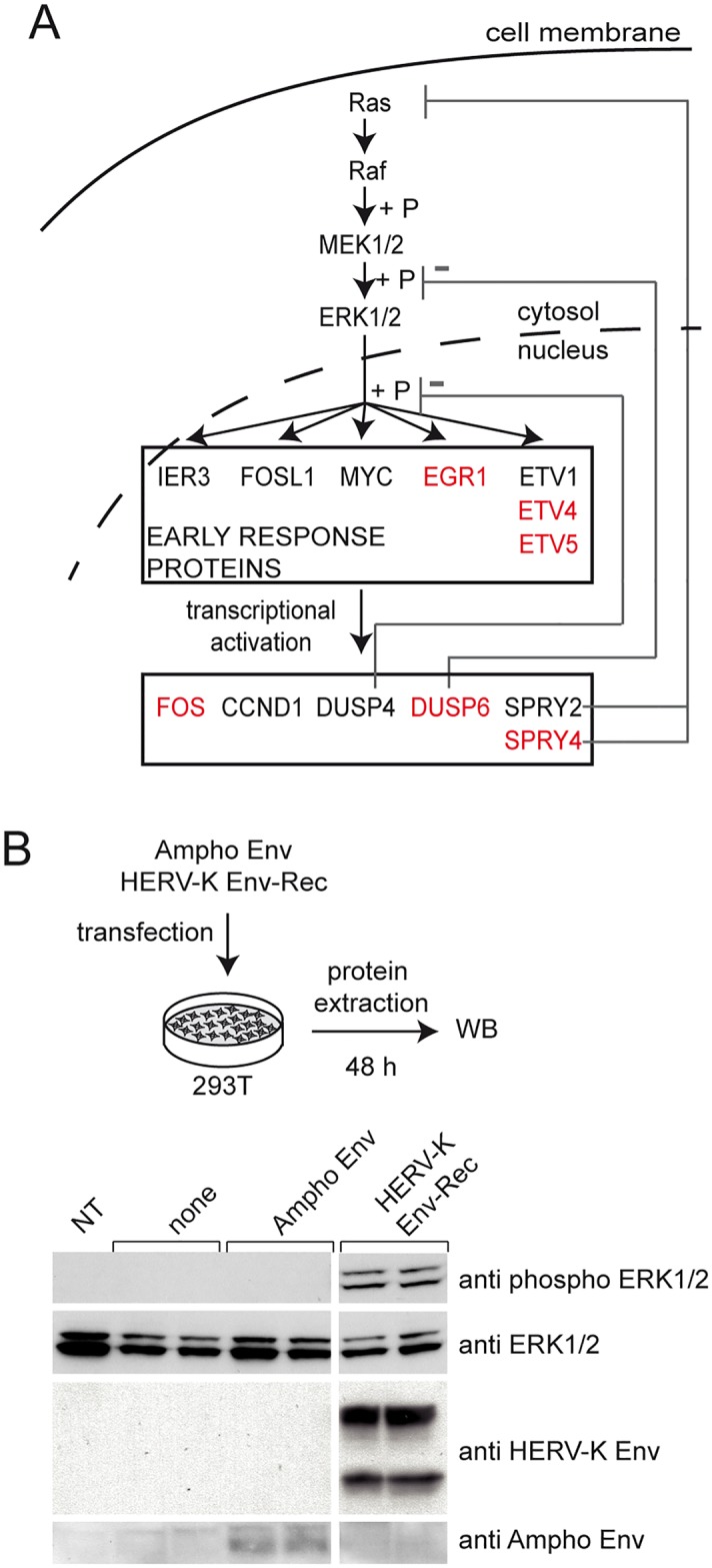
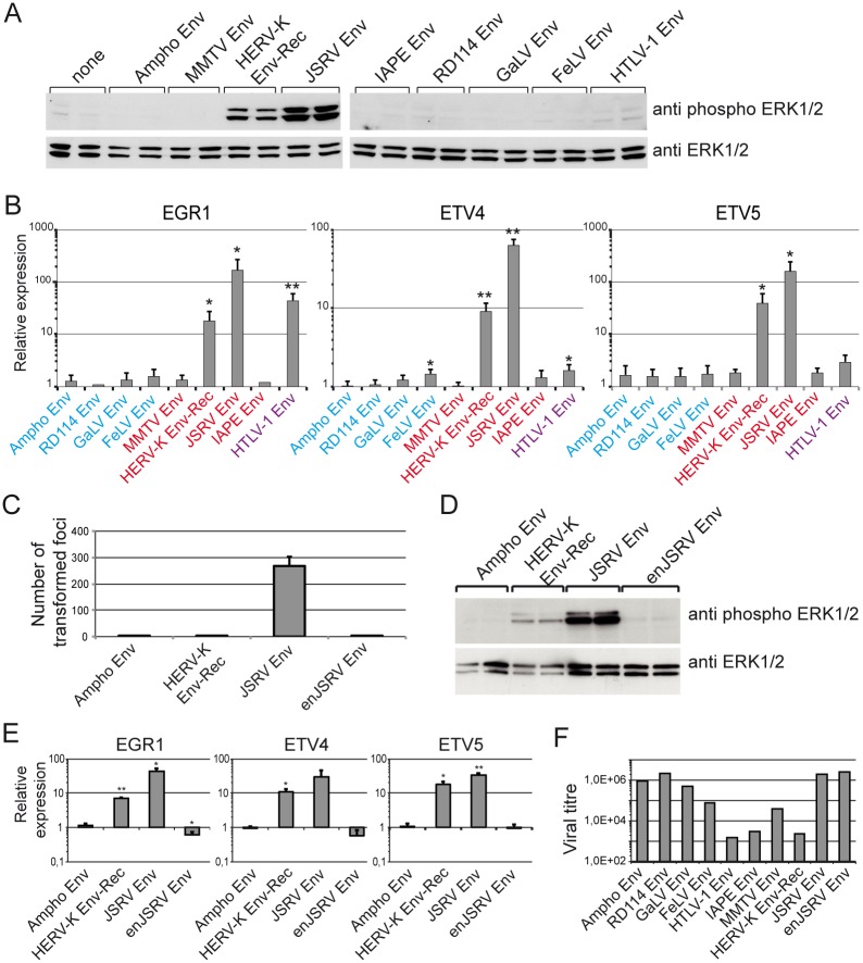
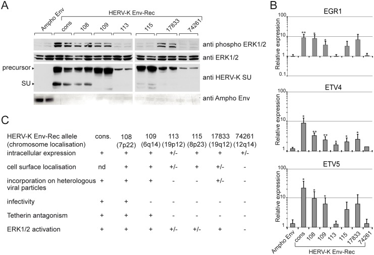
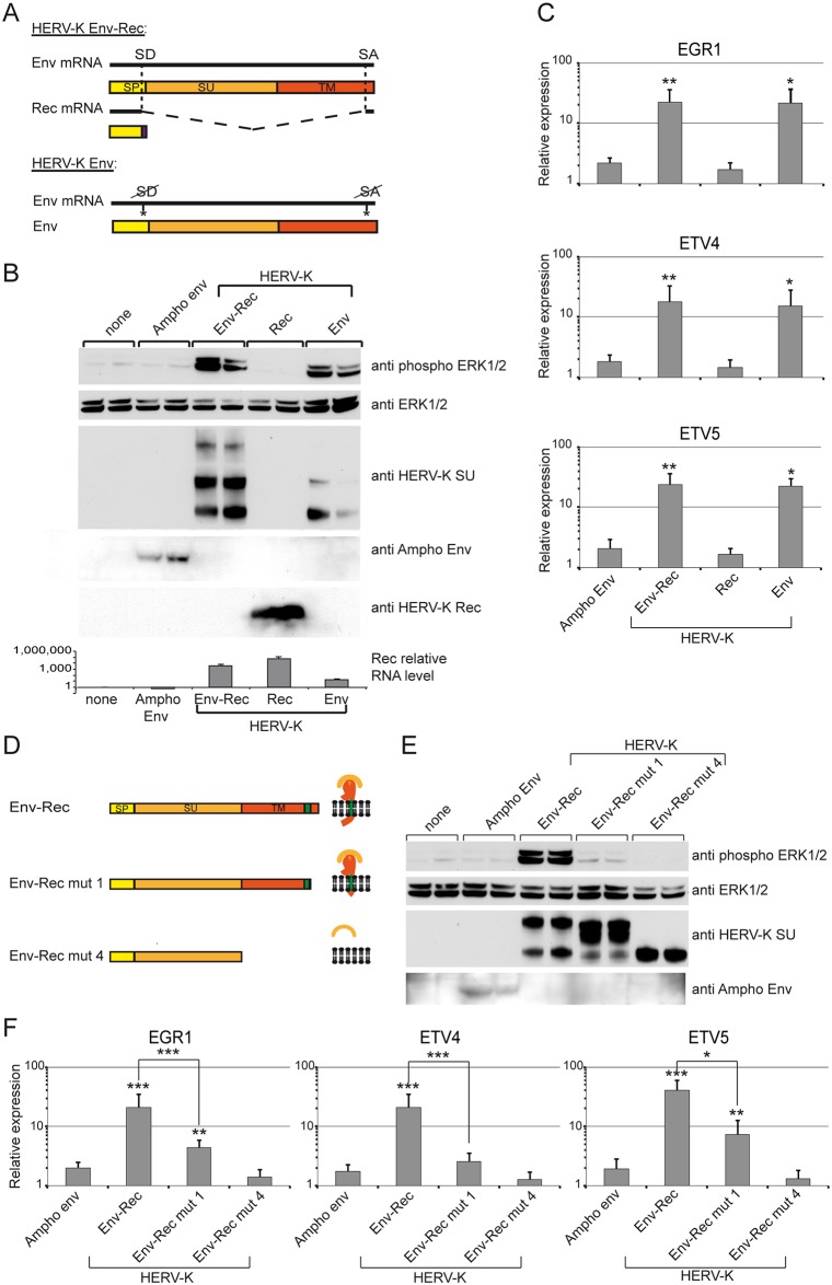
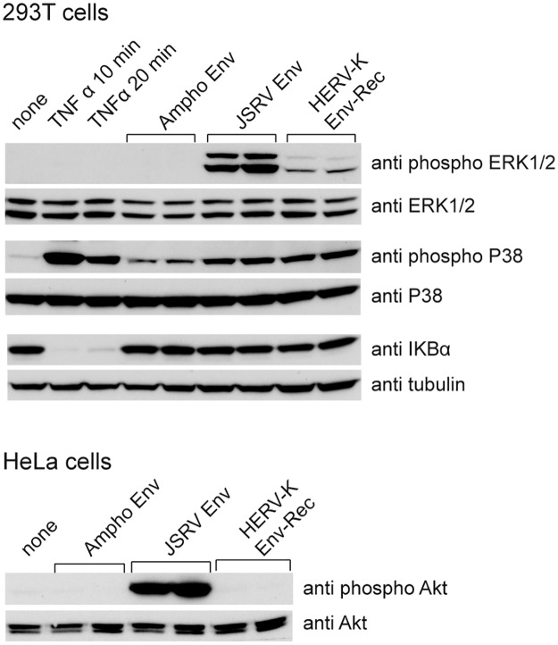
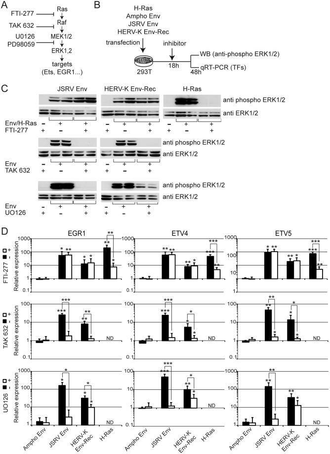
Endogenous retroviruses are cellular genes of retroviral origin captured by their host during the course of evolution and represent around 8% of the human genome. Although most are defective and transcriptionally silenced, some are still able to generate retroviral-like particles and proteins. Among these, the HERV-K(HML2) family is remarkable since its members have amplified relatively recently and many of them still have full length coding genes. Furthermore, they are induced in cancers, especially in melanoma, breast cancer and germ cell tumours, where viral particles, as well as the envelope protein (Env), can be detected. Here we show that HERV-K(HML2) Env per se has oncogenic properties. Its expression in a non-tumourigenic human breast epithelial cell line induces epithelial to mesenchymal transition (EMT), often associated with tumour aggressiveness and metastasis. In our model, this is typified by key modifications in a set of molecular markers, changes in cell morphology and enhanced cell motility. Remarkably, microarrays performed in 293T cells reveal that HERV-K(HML2) Env is a strong inducer of several transcription factors, namely ETV4, ETV5 and EGR1, which are downstream effectors of the MAPK ERK1/2 and are associated with cellular transformation. We demonstrate that HERV-K(HML2) Env effectively activates the ERK1/2 pathway in our experimental setting and that this activation depends on the Env cytoplasmic tail. In addition, this phenomenon is very specific, being absent with every other retroviral Env tested, except for Jaagsiekte Sheep Retrovirus (JSRV) Env, which is already known to have transforming properties in vivo. Though HERV-K Env is not directly transforming by itself, the newly discovered properties of this protein may contribute to oncogenesis.


Conflict of interest statement

The authors have declared that no competing interests exist.

Figures

Fig 1
Fig 1. HERV-K envelope protein induces EMT in MCF10A human breast epithelial cell line.
MCF10A cells were transduced with either a control “empty” vector (EV) or a vector expressing HERV-K Env-Rec. (A) Fluorescence microscopy images of MCF10A cell populations after selection with hygromycin compared to control wildtype cells (WT) and cells stimulated with 5ng/ml TGFβ (TGFβ). Images were taken at 10x magnification after staining the cells with Cell Tracker Green. (B) Changes in cell morphology were quantified by calculating the average cell size (area) of each population (a minimum of 1000 cells were counted for each population). (C) The expression levels of EMT related transcripts were evaluated in the different MCF10A populations by qRT-PCR. Genes of interest were normalized to the RPLO gene and expressed relative to WT cells. (D+E) The migration and invasion capacities of the cells were assessed using transwell assays. The cells were allowed to migrate and invade (through a layer of Matrigel) for 22 hours. DAPI staining was performed and cells that had migrated/invaded were observed under a fluorescent microscope and counted. Migration and invasion of TGF stimulated cells and HERV-K Env expressing cells is represented relative to WT and EV cells respectively. Error bars represent the standard deviation of at least 3 independently generated cell populations with triplicate readings taken from each population. Significant differences between cell populations were assessed using a one-tailed t test performed relative to WT cells (C+E) and EV cells (D); * = p<0.05; ** = p<0.01; *** = p<0.001.
Fig 2
Fig 2. HERV-K envelope protein induces expression of several transcription factors.
(A) Scheme of the experimental procedure. 293T cells were transfected with the indicated plasmids, in duplicate for each condition. 24 and 48 hours post transfection, cells were harvested for RNA extraction. Gene expression analysis was performed with the Agilent SurePrint G3 Human GE 8x60K Microarray. (B) Data from microarrays were processed and normalized as described in the material and methods in order to assess differentially expressed genes between control and HERV-K Env expressing samples. The genes listed here were from the 48 hour time point and selected by the following criteria: fold-change ≥2 and a false detection rate (FDR) <0.05. Expression levels of the top five genes were confirmed by qRT-PCR performed on the same samples. The results after normalisation with RPLO gene are indicated in the right column. (C) qRT-PCR was performed on total RNA extracted from Ampho Env (control) or HERV-K Env-Rec expressing cells at 48 hours post-transfection. The amount of mRNA transcripts for the indicated transcription factors (EGR1: Early Growth Response 1; ETV4 and ETV5: Ets variant 4/5) were normalised to the RPLO gene and expressed relative to non-transfected 293T cells. Error bars represent the standard deviation of five independent experiments. Significant differences in gene expression were assessed using a one-tailed t test performed relative to the Ampho Env condition.
Fig 3
Fig 3. HERV-K Env activates the ERK1/2 MAPK.
(A) Overview of the ERK1/2 MAPK pathway (adapted from [46]). A broad range of extracellular stimuli can activate the ERK1/2 pathway which starts with the activation of Ras. Activated Ras in turn activates Raf, the first kinase in the pathway. Sequential phosphorylation (shown by a +P) and activation of downstream kinases MEK1/2 subsequently activates ERK1/2 which can then translocate to the cell nucleus where it phosphorylates early response targets (including EGR1, ETV4 and ETV5). These targets act as transcriptional activators of several genes. Genes identified in the microarray data presented in Fig 2B are highlighted in red. (B) Activation of ERK1/2 was directly tested in 293T cells transfected with HERV-K Env-Rec (or indicated control proteins). Western Blots were performed on cell lysates harvested 48 hours post transfection and membranes were probed for phosphorylated ERK1/2, stripped and stained again for the total form of the kinases. (NT: non transfected). Expression of both HERV-K and Ampho Envs was also confirmed.
Fig 4
Fig 4. Oncogenic properties carried by different retroviral Envs.
(A) Phosphorylation of ERK1/2 in 293T cells expressing a panel of retroviral Envs was assayed as described in Fig 3B. Samples in left and right panels were prepared and processed (migration, transfer, revelation) at the same time; therefore intensity of the signals in both sets of panels can be compared directly. The Envs tested span three different retroviral genera and are colour-coded according to group (red: betaretroviruses; blue: gammaretrovirus; purple: deltaretrovirus). (B) The mRNA levels of the transcription factors EGR1, ETV4 and ETV5 following expression of the indicated Envs were measured as described in Fig 2C. Error bars represent standard deviation of the mean of three independent experiments. (C) The transforming activity of Ampho, HERV-K, JSRV and enJSRV retroviral Env proteins was assessed in 208F cells. Briefly, cells were transiently transfected with expression vectors for the 4 Env proteins and left to reach confluence. The number of transformed foci was counted after 3–4 weeks. The histogram represents the average number of foci obtained per 2x10⁵ cells in 3 independent experiments. (D) Phosphorylation of ERK1/2 in 293T cells expressing the 4 retroviral Envs mentioned above was assessed as in Fig 3B. (E) The mRNA levels of transfection factors EGR1, ETV4 and ETV5 following expression of the 4 Envs was tested as described in Fig 2C. (F) We ensured that all the expression vectors for the Env proteins were functional by measuring the viral titres obtained with them when pseudotyping heterologous retroviral particles. Ampho, RD114, GaLV, FelV and MMTV Envs were tested using a MLV core, HERV-K, JSRV, enJSRV, IAP and HTLV1 Envs were tested with a HIV core. Viral titres were measured on 293T cells, except for FeLV pseudotypes (feline G355.5 cells) and MMTV pseudotypes (mouse NIH 3T3 cells). Background infection levels (i.e. the titres obtained with no Env expression vector) were less than 100 whatever the core/target cells combination.
Fig 5
Fig 5. Test for ERK1/2 phosphorylation and expression of transcription factors induced by endogenous HERV-K Env alleles.
(A) Previously described endogenous alleles of HERV-K Env were tested for their ability to induce phosphorylation of ERK1/2 by Western blot on 293T cell lysates following transient transfection. Expression of each HERV-K Env allele was also assessed. We also checked that Ampho Env was properly expressed. (B) Expression levels of the transcription factors EGR1, ETV4 and ETV5 induced by the endogenous alleles of HERV-K Env were measured as described in Fig 2C. Error bars represent the standard deviation of the mean of four independent experiments. (C) Recapitulation of the functional properties of all HERV-K Env-Rec variants which have been assessed [43, 61] (nd: not determined).
Fig 6
Fig 6. Mapping of the domains involved in HERV-K Env-Rec activation of the ERK1/2 pathway and induction of the transcription factors.
(A) The HERV-K Env-Rec plasmid expresses two distinct proteins via alternative splicing: the full-length RNA encodes Env, a glycoprotein that is cleaved into two subunits during synthesis (surface subunit, SU, and transmembrane subunit, TM), while internal splicing sites (SD for splice donor and SA for splice acceptor) lead to the production of the Rec accessory protein. Introducing silent mutations in the splice sites generated an expression vector that expresses Env alone. (B) The different constructs were assayed for their capacity to activate the MAPK ERK1/2 as described in Fig 3B. The expression levels of Ampho Env, HERV-K Env and HERV-K Rec were also checked by Western blot. In the case of Rec, we also performed qRT-PCR reactions on RNA samples to detect specifically the spliced Rec transcripts. (C) Transcription factor expression following transfection of the constructs above was measured as described in Fig 2C. Error bars represent the standard deviation of five independent experiments. (D) Truncation mutants of HERV-K Env were generated to identify the region required for activation of the ERK1/2 pathway. The expected protein structure of each mutant is shown on the right. All modifications were designed to change Env without altering the Rec ORF and keeping the global structure of the Env-Rec RNA. The different domains of each construct are indicated: SP (signal peptide, yellow), SU (orange), TM (red). Env-Rec mut1 corresponds to a C-terminal truncated version of the complete envelope protein. Env-Rec mut4 is the soluble surface subunit. Expression of Ampho Env and the different HERV-K Env-Rec mutants was measured together with their ability to induce phosphorylation of ERK1/2 by Western Blot (E) and expression of the transcription factors was measured by qRT-PCR (F). Error bars represent the standard deviation of the mean of eight independent experiments.
Fig 7
Fig 7. HERV-K Env activation of other signalling pathways.
(Top panel) 293T cells were transfected by the indicated plasmids as described in Fig 3B, or stimulated with TNFα (100 ng/μL for 10 and 20 minutes), a positive control for activation of NFκB and p38 pathways. Lysates were tested by Western blot for phosphorylated and total (after membrane stripping) forms of the kinases ERK1/2 and p38, or for IκBα (for IκBα, tubulin was used as a control for homogenous loading). (Bottom panel) HeLa cells were transiently transfected by the indicated plasmids. Cell lysates were harvested 48 hours post transfection and phosphorylated and total (after membrane stripping) levels of AKT were assessed by western blotting.
Fig 8
Fig 8. HERV-K Env activates the ERK1/2 MAPK pathway upstream of Raf.
(A) The ERK1/2 pathway was targeted by different chemical inhibitors: FTI-277 (a H/K-Ras inhibitor), TAK 632 (a pan Raf inhibitor), and U0126 (a broadly-used antagonist of MEK1/2). (B) The experimental procedure is illustrated on the scheme. Briefly, 293T cells were transfected with the indicated plasmids. Inhibitors (or vehicle control) were added when media was changed 18 hours post-transfection. Lysates and total RNA were harvested 48 hours post transfection. Activation of ERK1/2 (C) and induction of transcription factor expression (D) were assayed as described previously (Figs 2C & 3B) in the presence (+) or absence (-) of FTI-277, TAK 632 or U0126. Error bars represent the standard deviation of the mean of five independent experiments.

Similar articles

Cited by

References

    1. Hofacre A, Fan H. Jaagsiekte sheep retrovirus biology and oncogenesis. Viruses. 2010. December;2(12):2618–48. doi: 10.3390/v2122618 - DOI - PMC - PubMed
    1. Caporale M, Cousens C, Centorame P, Pinoni C, De las Heras M, Palmarini M. Expression of the jaagsiekte sheep retrovirus envelope glycoprotein is sufficient to induce lung tumors in sheep. Journal of virology. 2006;80:8030–7. doi: 10.1128/JVI.00474-06 - DOI - PMC - PubMed
    1. Maeda N, Palmarini M, Murgia C, Fan H. Direct transformation of rodent fibroblasts by jaagsiekte sheep retrovirus DNA. Proc Natl Acad Sci U S A. 2001;98(8):4449–54. doi: 10.1073/pnas.071547598 - DOI - PMC - PubMed
    1. Allen TE, Sherrill KJ, Crispell SM, Perrott MR, Carlson JO, DeMartini JC. The Jaagsiekte sheep retrovirus envelope gene induces transformation of the avian fibroblast cell line DF-1 but does not require a conserved SH2 binding domain. J Gen Virol. 2002;83(Pt 11):2733–42. doi: 10.1099/0022-1317-83-11-2733 - DOI - PubMed
    1. Danilkovitch-Miagkova A, Duh FM, Kuzmin I, Angeloni D, Liu SL, Miller AD, et al. Hyaluronidase 2 negatively regulates RON receptor tyrosine kinase and mediates transformation of epithelial cells by Jaagsiekte sheep retrovirus. Proc Natl Acad Sci U S A. 2003;100(8):4580–5. doi: 10.1073/pnas.0837136100 - DOI - PMC - PubMed

MeSH terms

Substances

LinkOut - more resources