. 2017 Jun 26;13(6):e1006437.
doi: 10.1371/journal.ppat.1006437. eCollection 2017 Jun.

Macrophages, but not neutrophils, are critical for proliferation of Burkholderia cenocepacia and ensuing host-damaging inflammation

Affiliations

Macrophages, but not neutrophils, are critical for proliferation of Burkholderia cenocepacia and ensuing host-damaging inflammation

Jennifer Mesureur et al. PLoS Pathog. .

Erratum in

Abstract

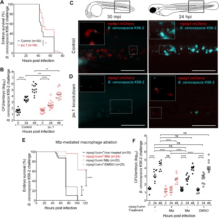
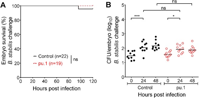
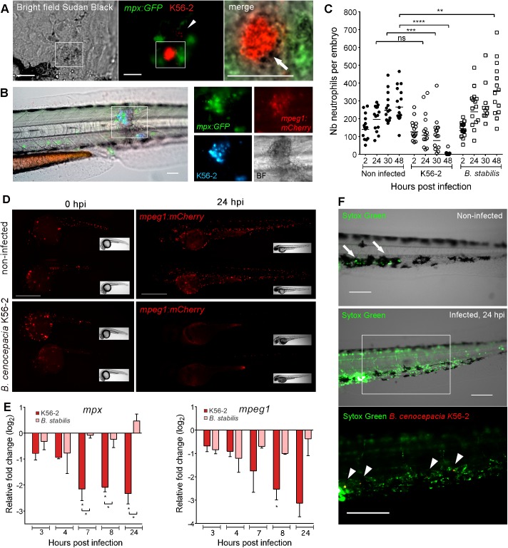
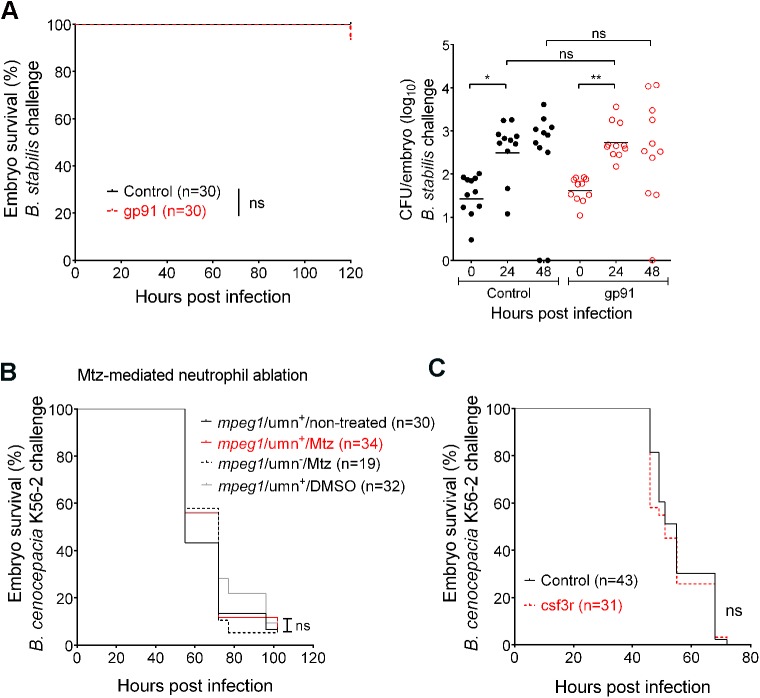
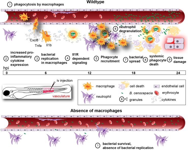
Bacteria of the Burkholderia cepacia complex (Bcc) can cause devastating pulmonary infections in cystic fibrosis (CF) patients, yet the precise mechanisms underlying inflammation, recurrent exacerbations and transition from chronic stages to acute infection and septicemia are not known. Bcc bacteria are generally believed to have a predominant extracellular biofilm life style in infected CF lungs, similar to Pseudomonas aeruginosa, but this has been challenged by clinical observations which show Bcc bacteria predominantly in macrophages. More recently, Bcc bacteria have emerged in nosocomial infections of patients hospitalized for reasons unrelated to CF. Research has abundantly shown that Bcc bacteria can survive and replicate in mammalian cells in vitro, yet the importance of an intracellular life style during infection in humans is unknown. Here we studied the contribution of innate immune cell types to fatal pro-inflammatory infection caused by B. cenocepacia using zebrafish larvae. In strong contrast to the usual protective role for macrophages against microbes, our results show that these phagocytes significantly worsen disease outcome. We provide new insight that macrophages are critical for multiplication of B. cenocepacia in the host and for development of a fatal, pro-inflammatory response that partially depends on Il1-signalling. In contrast, neutrophils did not significantly contribute to disease outcome. In subcutaneous infections that are dominated by neutrophil-driven phagocytosis, the absence of a functional NADPH oxidase complex resulted in a small but measurably higher increase in bacterial growth suggesting the oxidative burst helps limit bacterial multiplication; however, neutrophils were unable to clear the bacteria. We suggest that paradigm-changing approaches are needed for development of novel antimicrobials to efficiently disarm intracellular bacteria of this group of highly persistent, opportunistic pathogens.


Conflict of interest statement

The authors have declared that no competing interests exist.

Figures

Fig 1
Fig 1. Macrophages are critical for virulence of B. cenocepacia.
(A,B) Embryo survival (average inoculum 17 CFU, representative experiment) (A) and bacterial burden (total of 3 experiments) over time (B) of control (black) and pu.1 knockdown embryos (red) injected iv with B. cenocepacia K56-2. (C,D) Representative fluorescence overlay images of an mpeg1:mCherry control and mpeg1:mCherry pu.1 knockdown embryo at 30 min and 24 h after injection with ~40 CFU B. cenocepacia K56-2 (blue). See also S2 Fig. (C) mCherry-positive macrophages (red) colocalise with K56-2 at 30 mpi, and are no longer detected at 24 hpi (insets show magnification). (D) mCherry-positive macrophages are absent in knockdown embryos at 30 mpi and start to re-appear at 24 hpi (insets show magnification). Scale bars, 100 μm. (E,F) Embryo survival (average inoculum 28 CFU, representative experiment) (E) and corresponding bacterial burden (n = 10 per group per time point). (F) of mpeg1/umn+ embryos, untreated or treated with 5mM Mtz or 0.2% DMSO, and mpeg1/umnembryos treated with 5mM Mtz iv injected with B. cenocepacia K56-2. (B,F) Geometric means with each data point representing an individual embryo. Dead embryos marked as black open circles (not recorded for 5 embryos in (B), and in (F)). (A, B, E, F) * p ≤ 0.05, ** p ≤ 0.01, *** p ≤ 0.001; **** p ≤ 0.0001. ns: not significant. See materials and methods for statistical tests. See also S1 Fig and S3 Fig.
Fig 2
Fig 2. Macrophages clear less virulent Bcc strains inefficiently.
(A,B) Embryo survival (average inoculum 23 CFU, representative experiment) (A) and bacterial burden (total of 2 experiments) over time (B) of control (black) and pu.1 knockdown embryos (red) iv injected with B. stabilis LMG14294. Geometric means with each data point representing an individual embryo. * p ≤ 0.05, *** p ≤ 0.001; ns: not significant. See materials and methods for statistical tests. See also S1 Fig.
Fig 3
Fig 3. Acute, but not persistent infection results in systemic phagocyte death.
(A) Sudan black staining of an mpx:GFP embryo (24 hpi), injected with ~45 CFU B. cenocepacia K56-2. Bright field, fluorescence and merged images showing recruited neutrophils (green) that release granules (stained by Sudan black as black deposit, white arrow) close to an infected cell containing red fluorescent bacteria. Arrow head, individual bacteria. Scale bars, 50 μm. (B) Image of the trunk region of an mpx:GFP; mpeg1:mCherry embryo 24 h post iv injection in the blood island with B. cenocepacia K56-2 (Turquoise), showing neutrophils (green) and macrophages (red) infiltrated in an infection site with multiple infected cells. BF, Bright field image, showing tissue damage. Scale bar, 50 μm. (C) Mean neutrophil numbers in non-infected control and mpx:GFP embryos injected at 50 hpf with B. cenocepacia K56-2 or B. stabilis LMG14294. See also S5B Fig and S5C Fig. (D) Mpeg1:mCherry embryos showing reduced macrophage numbers (red) at 24 hpi in B. cenocepacia K56-2-infected (~45 CFU) compared to non-infected control embryos. Scale bars, 0.5 mm. See S5D Fig for quantification. (E) Mean relative mpx and mpeg1 gene expression level (qRT-PCR) in embryos injected with on average 234 CFU of B. cenocepacia K56-2 (red bars) or 123 CFU of B. stabilis LMG14294 (pink bars) each normalised to a PBS-injected control group at each time point and analysed using Anova (error bars, SEM). Two independent experiments. Asterisks below each bar indicate significance compared to the PBS control at each time point, significance between groups per time point is indicated with a horizontal line. (F) Non-infected and B. cenocepacia K56-2 (~50 CFU, Turquoise indicated in red for better visualization) infected embryos at 24hpi with the cell-impermeable dye Sytox Green. Arrows, dead cells due to DMSO injection. Arrow heads, bacterial clusters. Scale bars, 100 μm. See also S5F Fig. (C). Each data point represents an individual embryo. (C,E) * p ≤ 0.05; ** p ≤ 0.01; *** p ≤ 0.001; **** p ≤ 0.0001; ns: non-significant. See materials and methods for statistical tests. See also S5 Fig.
Fig 4
Fig 4. Neutrophils do not contribute significantly to infection.
(A) Embryo survival (left; representative experiment) and bacterial burden (total of 2 experiments) over time (right panel, geometric mean) of control (black) and gp91 knockdown embryos (red) iv injected with B. stabilis. Each data point represents an individual embryo. (B) Embryo survival of mpx/umn+ embryos, untreated or treated with 10 mM Mtz or 0.2% DMSO, and mpx/umnembryos treated with 10 mM Mtz iv injected with ~ 50 CFU B. cenocepacia K56-2. (C) Embryo survival (average inoculum 50 CFU, representative experiment) of control (black) and csf3R knockdown embryos (red) injected iv with B. cenocepacia K56-2. * p ≤ 0.05; ** p ≤ 0.01; ns: non-significant. See materials and methods for statistical tests.
Fig 5
Fig 5. Neutrophils efficiently phagocytose surface-associated Bcc bacteria.
Confocal stacks after subcutaneous infection with B. stabilis in mpx:GFP embryos (STD intensity projection, 2 μm x 19 steps; T = 35–37 in S2 Movie). Arrows, rounded neutrophils (green) with vacuoles full of bacteria (red) at 64 minutes post injection (mpi) eject their cell contents in the surroundings (arrow heads 65.5 mpi), leaving bacterial clusters and cell debris (arrow heads, 66 min). Lower panels, MAX intensity projection of three consecutive slices (2 μm) at 65.5 mpi showing ejected cellular contents (diffuse GFP signal). Scale bars, 50 μm.
Fig 6
Fig 6. Macrophages, but not neutrophils, contribute to increased bacterial burden and pro-inflammatory responses towards subcutaneously introduced B. cenocepacia.
(A) Mpeg1/umn+ embryos were subcutaneously injected with B. cenocepacia K56-2 expressing Turquoise. Fluorescent overlay images were taken at 90 min and 5 h post infection, showing infected macrophages (red). Scale bars, 10 μm. (B) Images (red and blue overlay and below slightly enlarged individual fluorescence images with blue filter) of the indicated area (see drawing) of an mpx/umn+ (B1) and an mpeg1/umn+ (B2) embryo subcutaneously injected with B. cenocepacia K56-2 expressing Turquoise. B1 shows an embryo followed in time displaying neutrophil infiltration (red) and increase in bacterial burden (blue, see Fig 7A for quantification). B2 shows the image of an embryo with macrophage infiltration (red) and high bacterial burden at 24 hpi. Arrow head points at mCherry positive debris. See non-infected mpx/umn+ and mpeg1/umn+ control embryos in S6 Fig for comparison. Scale bars, 100 μm. (C) Image of the infected area of a representative mpx/umn+ embryo depleted of neutrophils with Mtz, and subcutaneously injected with B. cenocepacia K56-2 (Turquoise) at 24 hpi. Scale bar 100 μm, and 50 μm for inset. See also S6D Fig. (D) Image of a representative mpeg1/umn+ embryo depleted of macrophages with Mtz and subcutaneously injected with B. cenocepacia K56-2 (Turquoise) at 24 hpi. Scale bar 100 μm, and 50 μm for inset.
Fig 7
Fig 7. The absence of Gp91 results in increased bacterial burden and neutrophil persistence during subcutaneous infections.
(A) Bacterial burden over time after subcutaneous injection of B. cenocepacia K56-2 in control (black circles) and gp91 knockdown embryos (open red circles). Average of two independent experiments. (B) Bacterial burden over time after subcutaneous injection of B. stabilis LMG14294 in control (black circles) and gp91 knockdown embryos (open red circles). Average of three independent experiments. (C) Fluorescent overlay images of the injected area of a representative mpx:GFP control MO and gp91 knockdown embryo (neutrophils in green) in time after subcutaneous injection with B. stabilis (red). Inset shows bacterial load at ~20 hpi. % at 22 and 21 hpi indicates percentage of control and gp91 knockdown embryos that show reduced neutrophil numbers (86.8%), and persistent neutrophil infiltration (71.3%), respectively, at the infection site. Scale bar, 100 μm. (A,B) * p ≤ 0.05; **** p ≤ 0.0001; ns: non-significant. See materials and methods for statistical tests.
Fig 8
Fig 8. B. cenocepacia K56-2 induces robust pro-inflammatory Il1b expression that is dependent on macrophages.
(A,B) Mean relative il1b (A) and cxcl8 (B) gene expression levels (qRT-PCR) in embryos injected with on average 250 CFU B. cenocepacia K56-2 (red bars) or on average 111 CFU B. stabilis LMG14294 (pink bars), normalized to a PBS-injected control group at each time point. Error bars represent mean with SEM of three biological replicates. Asterisks above each bar indicate significance compared to the PBS control at each time point, significance between groups per time point is indicated with a horizontal line. (C,D) mpeg1/umn+ embryos were pre-treated at 34 hpf for 15 h with DMSO or 5 mM Mtz. Randomized groups were injected with either PBS or with B. cenocepacia K56-2 (on average 150 CFU). Graphs show mean relative il1b (C) and cxcl8 (D) gene expression levels (qRT-PCR) normalized to the PBS-injected DMSO-treated group at each time point. Error bars represent mean with SEM of two biological replicates. See also S8 Fig. (E) Confocal stack images (green/red overlay (left panels) and red channel (right panels)) of il1b:GFP/mpeg1:mCherry embryos 8 h post iv injection with PBS, or B. cenocepacia K56-2 (DS-Red). Due to strong fluorescence of GFP in epithelial cells in the trunk and head region, images were taken over the yolk sac valley. Scale bars 10 μm. (F) Embryo survival (average inoculum 44 CFU, representative experiment) of control (black) and il1b knockdown embryos (red) injected iv with B. cenocepacia K56-2. (G,H) Embryo survival (G) and bacterial burden over time (H) of control (black) and Anakinra-treated embryos (red) injected iv with B. cenocepacia K56-2 (average inoculum 107 CFU for both groups). Representative experiment. (A-D, F-H) * p ≤ 0.05; ** p ≤ 0.01; *** p ≤ 0.001; **** p ≤ 0.0001; ns: non-significant.
Fig 9
Fig 9. Schematic showing the role of macrophages during acute infection by B. cenocepacia K56-2 in zebrafish embryos.
Macrophages are the major phagocytosing cells of iv injected bacteria (1) for which they provide a critical replication niche (3). In their absence, K56-2 does not replicate (bottom panel, 1). Intracellular bacteria induce a rapid and robust increase in pro-inflammatory cytokine expression (2). Il1 signalling contributes to fatal pro-inflammatory responses (4), characterized by massive neutrophil and macrophage infiltration (5) neutrophil degranulation (6), bacterial dissemination from infected cells (7), systemic phagocyte death (8), and tissue damage (9).

Comment in

  • Macrophages as drivers of an opportunistic infection.
    Vergunst AC, Carranza NL, Zhang L, Gomes MC, Tasrini Y, Meijer AH, O'Callaghan D. Vergunst AC, et al. Microb Cell. 2017 Sep 13;4(10):362-364. doi: 10.15698/mic2017.10.595. Microb Cell. 2017. PMID: 29082233 Free PMC article.

References

    1. Drevinek P, Mahenthiralingam E. Burkholderia cenocepacia in cystic fibrosis: epidemiology and molecular mechanisms of virulence. Clin Microbiol Infect. 2010; 16(7):821–30. doi: 10.1111/j.1469-0691.2010.03237.x - DOI - PubMed
    1. Medina-Pascual M, Valdezate S, Villalón P, Garrido N, Rubio V, Saéz-Nieto J. Identification, molecular characterisation and antimicrobial susceptibility of genomovars of the Burkholderia cepacia complex in Spain. Eur J Clin Microbiol Infect Dis. 2012;31:3385–96. doi: 10.1007/s10096-012-1707-6 - DOI - PubMed
    1. Katsiari M, Roussou Z, Tryfinopoulou K, Vatopoulos A, Platsouka E, Maguina A. Burkholderia cenocepacia bacteremia without respiratory colonization in an adult intensive care unit: epidemiological and molecular investigation of an outbreak. Hippokratia. 2012;16(4):317–23. - PMC - PubMed
    1. Bressler A, Kaye K, LiPuma J, Alexander B, Moore C, Reller L, et al. Risk factors for Burkholderia cepacia complex bacteremia among intensive care unit patients without Cystic Fibrosis: A case-control study. Infect Control Hosp Epidemiol. 2007;28(8):951–8. doi: 10.1086/519177 - DOI - PubMed
    1. Hartl D, Gaggar A, Bruscia E, Hector A, Marcos V, Jung A, et al. Innate immunity in cystic fibrosis lung disease. J Cyst Fibros. 2012; 11(5):363–82. doi: 10.1016/j.jcf.2012.07.003 - DOI - PubMed

MeSH terms