. 2017 Sep:509:205-221.
doi: 10.1016/j.virol.2017.06.008. Epub 2017 Jun 23.

Cytomegalovirus UL128 homolog mutants that form a pentameric complex produce virus with impaired epithelial and trophoblast cell tropism and altered pathogenicity in the guinea pig

Affiliations

Cytomegalovirus UL128 homolog mutants that form a pentameric complex produce virus with impaired epithelial and trophoblast cell tropism and altered pathogenicity in the guinea pig

Stewart Coleman et al. Virology. 2017 Sep.

Abstract

Guinea pig cytomegalovirus (GPCMV) encodes a homolog pentameric complex (PC) for specific cell tropism and congenital infection. In human cytomegalovirus, the PC is an important antibody neutralizing target and GPCMV studies will aid in the development of intervention strategies. Deletion mutants of the C-terminal domains of unique PC proteins (UL128, UL130 and UL131 homologs) were unable to form a PC in separate transient expression assays. Minor modifications to the UL128 homolog (GP129) C-terminal domain enabled PC formation but viruses encoding these mutants had altered tropism to renal and placental trophoblast cells. Mutation of the presumptive CC chemokine motif encoded by GP129 was investigated by alanine substitution of the CC motif (codons 26-27) and cysteines (codons 47 and 62). GP129 chemokine mutants formed PC but GP129 chemokine mutant viruses had reduced epitropism. A GP129 chemokine mutant virus pathogenicity study demonstrated reduced viral load to target organs but highly extended viremia.

Keywords: Congenital infection; Cytomegalovirus; Epithelial cells; Glycoproteins; Guinea pig; Pentameric complex; Placenta; Trophoblast; UL128; Viral tropism.


Figures

Figure 1
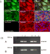
Figure 1. Establishment of a guinea pig trophoblast cell line
(i) Trophoblast cell lines express cytokeratin. Primary cells were clonally isolated from the guinea pig placenta and immortalized with HPV E6/E7 as described in materials and methods. Both primary (A–C) and transformed trophoblast cells lines (D–F), along with guinea pig lung fibroblast control (G–I), were characterised with antibodies directed against cytokeratin (secondary anti-mouse IgG FITC conjugate) (67) (A, D and G). Cells were counter-stained with F-actin stain, phalloidin 594 (B, E and H) and DAPI for cell nucleus (C, F and I) as described in materials and methods. (ii) RT-PCR assay for vimentin expression in primary and transformed trophoblasts. RT-PCR for vimentin (A) expression from RNA extracted from primary and transformed guinea pig trophoblast cells, guinea pig embryonic fibroblasts (GEF), and GPL cells using guinea pig specific vimentin primers (Table S1) or GAPDH (B). Primary trophoblast (lane 1), transformed trophoblasts (lane 2), GEF cells (lane 3), GPL cells (lane 4) and no template control (lane 5). RT-PCR assay studies were carried out as previously described and products analysed by agarose gel electrophoresis (67).
Figure 2
Figure 2. GPCMV encoding truncated GP129 is non-permissive for trophoblast cells
Trophoblast (D–E and J–L) and GPL (A–C and G–I) cell monolayers were infected with wt GPCMV (SG) or GP129 C-terminal deletion (102–179aa) mutant (NRD13) at moi of 1 pfu/cell. Cells stained by immunofluorescence assay with anti-IE2 antibody (A, D, G and J) to verify GPCMV infection and anti-pan cytokeratin antibody (B, E, H and K) to confirm epithelial cells as previously described (67). Cells were counterstained with DAPI (C, F, I and & L).
Figure 3
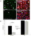
Figure 3. Bafilomycin A1 treatment inhibits SG GPCMV infection of trophoblast cells
GPL and Trophoblast cells were pre-treated with 50nM bafilomycin 1A prior to viral infection with SG GPCMV at moi of 1pfu/cell (67). (i) Immunofluorescence images of untreated (A and B) or bafilomycin pre-treated (C and D) trophoblast cells stained with anti-IE2 antibody (A and C) or anti-pan cytokeratin antibody and counterstained with DAPI (B and D). (ii) Percentage of infected trophoblast or GPL cells either untreated (black) or bafilomycin treated (grey) cells (ii). Virus detected by IE2 expression (i). Thirty random fields for 3 independent experiments per treatment were counted. Statistical analysis was performed with student t-test.
Figure 4
Figure 4. GP131 and GP133 C terminus mutants form a complex with other pentameric components
(i) Hopp/Woods hydrophilicity plot of GP131 predicted protein with deleted C-terminus sequence (121–192aa) to generate GP131d120 highlighted in red box. (ii) Immunoprecipitation assay of transient transfected epitope tagged pentameric components (gHGFP, gLmCherry, GP129MYC, GP133FLAG, and GP131d120MYC) with GFP-Trap (67). Western blot analysis of the immunoprecipitate using respective primary antibodies anti-GFP (gH); anti-mCherry (gL); anti-myc (GP131 d120MYC); anti-FLAG (GP133); and anti-myc (GP129). (iii) Hydrophilicity plot of GP133 predicted protein with deleted C-terminus sequence (76–127aa) to generate GP133d75 highlighted in red box. (iv) Western blot analysis of PC immunoprecipitation (gHGFP, gLmCherry, GP129MYC, GP131HA, and GP133d75FLAG) as previously described (67), using respective primary antibodies anti-GFP (gH); anti-mCherry (gL); anti-HA (GP131); anti-FLAG (GP133d75); and anti-myc (GP129). Western blot (ii) and (iv): Lane 1= Transfected cell lysate, Lane 2= unbound protein, Lane 3 = immunoprecipitation and Lane 4= untransfected cell lysate.
Figure 5
Figure 5. GP129 C-terminus deletion mutants form a complex with other pentameric components
(i) Hopp/Woods hydrophilicity plot of the wild-type GP129. The non-epithelial tropic GPCMV encodes a GP129 deletion from codon 102 – 179 (black dotted line). GP129 mutants with different sequence deletions are shown: GP129DEL1 (deleted codons 102- 120) (red box); GP129DEL2 (deleted codons 121–140) (blue box); GP129DEL3 (deleted codons 145–178) (green box). (ii) Protein sequence alignment of wt GP129 and GP129 C-terminus deletion mutants. BLAST protein sequence alignment of 100–179 amino acids of wt GP129, compared to the C-terminal GP129 mutants: GP129NRD13 (deletion 102–179); GP129DEL1 (deletion 102–120); GP129DEL2 (deletion 121–140); and GP129DEL3 (deletion 145–178). (iii) Immunoprecipitation assay of transient transfected epitope tagged pentameric components (gHGFP, gLmCherry, GP131HA, GP133FLAG) and myc tagged GP129 C-terminus mutant constructs (GP129DEL1, GP129DEL2, or GP129DEL3) using GFP-Trap (67). Western blot analysis of the immunoprecipitate using respective primary antibodies gH (anti-GFP); gL (anti-mCherry); GP131 (anti-HA); GP133 (anti-FLAG); and GP129 mutant constructs (anti-myc) and visualized with appropriate secondary antibody-HRP. (iv) Control western blot analysis of immunoprecipitation using GFP-Trap of pentameric complex components with GFP substituted for gHGFP. Western blot (iii) and (iv): Lane 1= Transfected cell lysate, Lane 2= unbound protein, Lane 3 = immunoprecipitation and Lane 4= untransfected cell lysate.
Figure 6
Figure 6. Schematic of parental viruses and modified mutant viruses
Schematic diagram of GPCMV genome (BAC and virus) with pentameric complex genes indicated. The native truncated GP129 is shown in red in GP129-133 locus. Ectopic expression of GP129 cDNA in the intergenic GP25-GP26 locus was as described in materials and methods. GP129 ORFs were under under SV40 promoter (red rectangle) and SV40 polyA termination sequence (blue rectangle). Insertion locus of BAC in the GPCMV genome indicated (green box). The BAC plasmid was excised upon generation of recombinant GPCMV via Cre recombinase. Viruses: (i) wild type virus (SG); (ii) NRD13, encoding truncated GP129; (iii) GP129FRT, encoding wt GP129 cDNA in the GP25/GP26 locus; (iv) ** vDEL1, vDEL2, vDEL3, encoding C-terminal mutant GP129 cDNA in the GP25/GP26 locus; (v) vdROX with native GP129 deleted (GP129-GP133 locus), (vi) vdROXGP129, encoding wt GP129 cDNA in the GP25/GP26 locus; (vii) ***vCC1d and vCC2d, encoding CXC motif mutant GP129 cDNA.
Figure 7
Figure 7. Growth kinetics of GPCMV GP129 C-terminal mutants on renal epithelial, trophoblast and fibroblast cells
Comparative growth curves for viruses on fibroblast and epithelial cell lines. Viruses: wt (SG) (black circle); GP129FRT (red diamond); vDEL1 (green triangle); vDEL2 (purple square); vDEL3 (blue square); NRD13 (yellow circle) and vGP133Km CRE GPCMV. Cell lines: GPL fibroblasts (i) and (ii); renal epithelial (iii); and trophoblast cells (iv). Input virus on all cell lines, moi=1pfu/cell. Virus yields are total cell culture virus.
Figure 8
Figure 8. GP129 chemokine mutants form a complex with other pentameric components
(i) Blast alignment of predicted protein sequence 1-90 amino acids of GP129 wt and GP129 chemokine mutants GP129CC1d and GP129CC2d. Chemokine mutant GP129CC1d substituted CC sequence (codons 22–23) with AA (red box). GP129CC2d substituted CC sequence (red box) and two C (codons 46 and 62) with A (blue boxes). The predicted signal peptide sequence is marked with black arrow, cleavage site between amino acids 15 and 16. (ii) Immunofluorescence visualization of renal epithelial cells transfected with wt GP129 (A–D) or mutants, GP129CC1d (E–H) or GP129CC2d (I–L). GP129 co-transfected with gHGFP (A, E and I) and gLmCherry (B, F and J), GP129 detected by anti-myc (anti-mouse IgG-Cy5 (67)). Cells were counterstained with DAPI (D and H). (iii) Western blot analysis of GFP-Trap immunoprecipitation assay on transiently transfected epitope tagged pentameric components and myc epitope tagged chemokine mutant GP129CC1d and GP129CC2d constructs. Detection of proteins with primary antibodies gH (anti-GFP); gL (anti-mCherry); GP131 (anti-HA); GP133 (anti-FLAG); GP129CC1d and GP129CC2d (anti-myc) and respective secondary HRP conjugated antibody as previously described (67). Lane 1= Transfected cell lysate, Lane 2= unbound protein, Lane 3 = immunoprecipitation and Lane 4= mock control.
Figure 9
Figure 9. Viral growth kinetics of GP129 chemokine mutant viruses on renal epithelial, trophoblast epithelial and fibroblast cells
Comparative growth curves for GPCMV SG (black circle), GP129FRT (red diamond), dROXGP129 (orange circle), GP129CC1d (blue triangle), GP129CC2d (green square), and dROX (purple diamond) on GPL (i), renal epithelial (ii) and trophoblast (iii) cell monolayers. Input moi=1pfu/cell. Virus yields are total cell culture virus.
Figure 10
Figure 10. Comparative pathogenicity study of GP129 chemokine mutant viruses with wild type virus in animal model
Five separate groups (n=12 per group) of animals were infected subcutaneously (106 pfu) with virus: wt (SG) GPCMV (black), GP129FRT (grey), dROXGP129 (green), NRD13 (red), or vCC2d (blue) viruses dependent upon group assignment. NRD13 encodes a truncated GP129 (102–178 codon deletion) and vCC2d is a GP129 CC chemokine motif mutant. At 4, 8, 12 and 27 days post infection (dpi), 3 animals per group were evaluated for viral load in target organs: Lung (i), Liver (ii), and Spleen (iii) by real time PCR of tissue extracted DNA (67). Viral load plotted as viral genome copies/mg tissue. Salivary gland tissue was only evaluated at 27 dpi (iv). Blood viremia at 4, 8, 12 and 27 dpi was plotted as genome copies/ml blood (v). Statistical analysis performed by analysis of variance (ANOVA) between groups. Statistical groups: a) p <0.0005; b) p < 0.05; c) non-significant.

Similar articles

Cited by

References

    1. Dolan A, Cunningham C, Hector RD, Hassan-Walker AF, Lee L, Addison C, Dargan DJ, McGeoch DJ, Gatherer D, Emery VC, Griffiths PD, Sinzger C, McSharry BP, Wilkinson GW, Davison AJ. Genetic content of wild-type human cytomegalovirus. J Gen Virol. 2004;85(Pt 5):1301–12. - PubMed
    1. Sinclair J, Sissons P. Latency and reactivation of human cytomegalovirus. J Gen Virol. 2006;87(Pt 7):1763–79. doi: 10.1099/vir.0.81891-0. - DOI - PubMed
    1. Ramanan P, Razonable RR. Cytomegalovirus infections in solid organ transplantation: a review. Infect Chemother. 2013;45(3):260–71. doi: 10.3947/ic.2013.45.3.260. - DOI - PMC - PubMed
    1. Kim S, Snider JJ, Gill MJ. Cytomegalovirus disease in HIV infection: twenty years of a regional population’s experience. Clin Infect Dis. 2006;42(12):1808–9. doi: 10.1086/504435. - DOI - PubMed
    1. Ross SA, Boppana SB. Congenital cytomegalovirus infection: outcome and diagnosis. Semin Pediatr Infect Dis. 2005;16(1):44–9. doi: 10.1053/j.spid.2004.09.011. - DOI - PubMed

Publication types

MeSH terms