

Layered smooth muscle cell-endothelial progenitor cell sheets derived from the bone marrow augment postinfarction ventricular function
- PMID: 28651946
- PMCID: PMC5947323
- DOI: 10.1016/j.jtcvs.2017.04.081
Layered smooth muscle cell-endothelial progenitor cell sheets derived from the bone marrow augment postinfarction ventricular function
Abstract
Objective: The angiogenic potential of endothelial progenitor cells (EPCs) may be limited by the absence of their natural biologic foundation, namely smooth muscle pericytes. We hypothesized that joint delivery of EPCs and smooth muscle cells (SMCs) in a novel, totally bone marrow-derived cell sheet will mimic the native architecture of a mature blood vessel and act as an angiogenic construct to limit post infarction ventricular remodeling.
Methods: Primary EPCs and mesenchymal stem cells were isolated from bone marrow of Wistar rats. Mesenchymal stem cells were transdifferentiated into SMCs by culture on fibronectin-coated culture dishes. Confluent SMCs topped with confluent EPCs were detached from an Upcell dish to create a SMC-EPC bi-level cell sheet. A rodent model of ischemic cardiomyopathy was then created by ligating the left anterior descending artery. Rats were randomized into 3 groups: cell sheet transplantation (n = 9), no treatment (n = 12), or sham surgery control (n = 7).
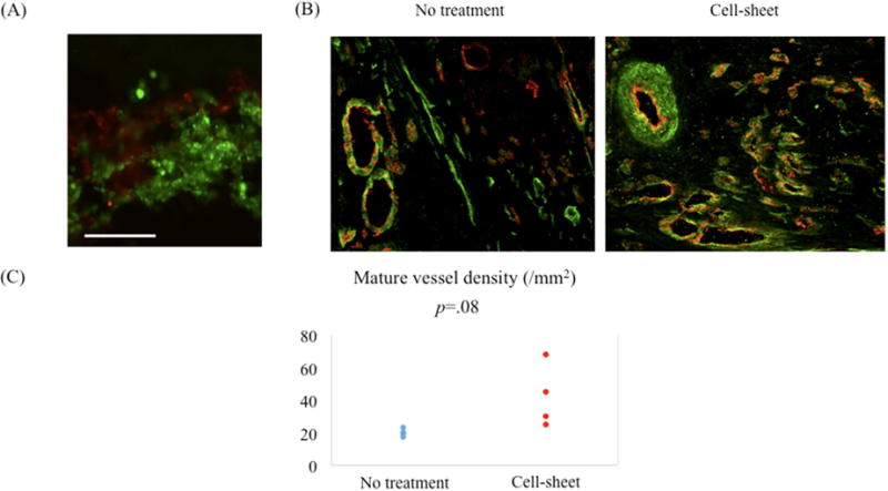
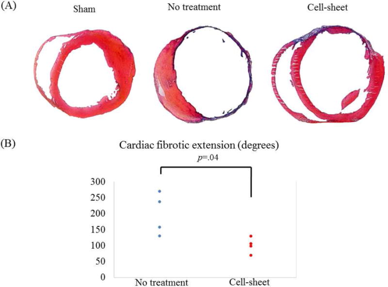
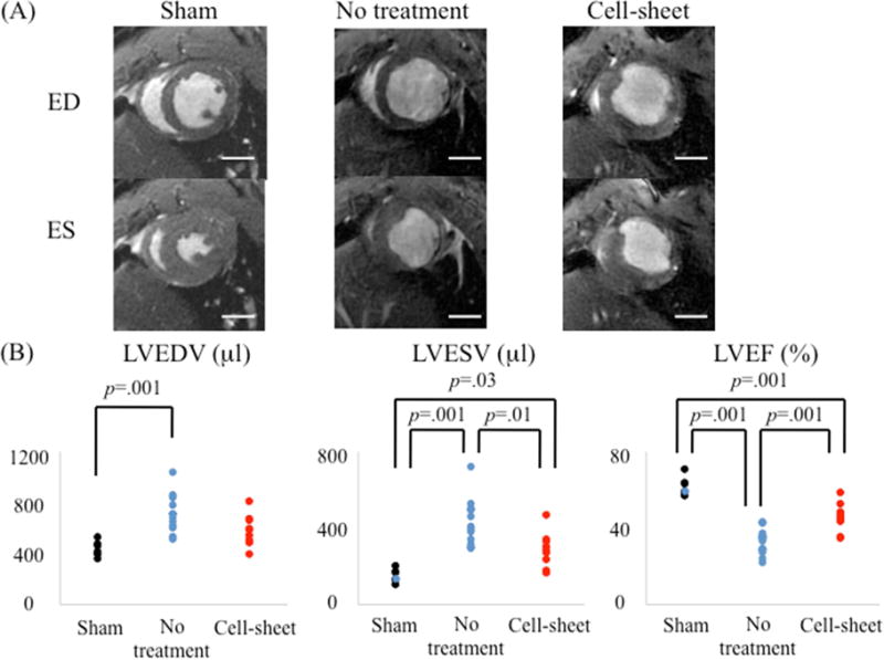
Results: Four weeks postinfarction, mature vessel density tended to increase in cell sheet-treated animals compared with controls. Cell sheet therapy significantly attenuated the extent of cardiac fibrosis compared with that of the untreated group (untreated vs cell sheet, 198 degrees [interquartile range (IQR), 151-246 degrees] vs 103 degrees [IQR, 92-113 degrees], P = .04). Furthermore, EPC-SMC cell sheet transplantation attenuated myocardial dysfunction, as evidenced by an increase in left ventricular ejection fraction (untreated vs cell sheet vs sham, 33.5% [IQR, 27.8%-35.7%] vs 45.9% [IQR, 43.6%-48.4%] vs 59.3% [IQR, 58.8%-63.5%], P = .001) and decreases in left ventricular dimensions.
Conclusions: The bone marrow-derived, spatially arranged SMC-EPC bi-level cell sheet is a novel, multilineage cellular therapy obtained from a translationally practical source. Interactions between SMCs and EPCs augment mature neovascularization, limit adverse remodeling, and improve ventricular function after myocardial infarction.
Keywords: cell sheet; myocardial infarction; neovascularization; regeneration; stem cells; tissue engineering.
Copyright © 2017 The American Association for Thoracic Surgery. Published by Elsevier Inc. All rights reserved.
Conflict of interest statement
Figures

Comment in
-
Recapitulating nature's design: Myocardial repair with cell sheet technology.J Thorac Cardiovasc Surg. 2017 Sep;154(3):951-952. doi: 10.1016/j.jtcvs.2017.05.011. Epub 2017 May 17. J Thorac Cardiovasc Surg. 2017. PMID: 28629840 No abstract available.
-
Beyond proof of concepts for ideal cardiac regenerative therapy.J Thorac Cardiovasc Surg. 2017 Sep;154(3):964-965. doi: 10.1016/j.jtcvs.2017.05.015. Epub 2017 May 17. J Thorac Cardiovasc Surg. 2017. PMID: 28645824 No abstract available.
-
Discussion.J Thorac Cardiovasc Surg. 2017 Sep;154(3):962-963. doi: 10.1016/j.jtcvs.2017.04.087. Epub 2017 Jun 23. J Thorac Cardiovasc Surg. 2017. PMID: 28651941 No abstract available.
References
-
- Dominici M, Le Blanc K, Mueller I, Slaper-Cortenbach I, Marini F, Krause D, et al. Minimal criteria for defining multipotent mesenchymal stromal cells. The International Society for Cellular Therapy position statement. Cytotherapy. 2006;8:315–317. - PubMed
-
- Thyberg J, Hedin U, Sjolund M, Palmberg L, Bottger BA. Regulation of differentiated properties and proliferation of arterial smooth muscle cells. Arteriosclerosis. 1990;10:966–990. - PubMed
-
- Langer R, Vacanti JP. Tissue engineering. Science. 1993;260:920–926. - PubMed
MeSH terms
Grants and funding
LinkOut - more resources
Full Text Sources
Other Literature Sources
Medical
