Case Reports
. 2017 Aug 15;77(16):4258-4267.
doi: 10.1158/0008-5472.CAN-17-1052. Epub 2017 Jun 26.

Unpaired Extracellular Cysteine Mutations of CSF3R Mediate Gain or Loss of Function

Affiliations
Case Reports

Unpaired Extracellular Cysteine Mutations of CSF3R Mediate Gain or Loss of Function

Haijiao Zhang et al. Cancer Res. .

Abstract

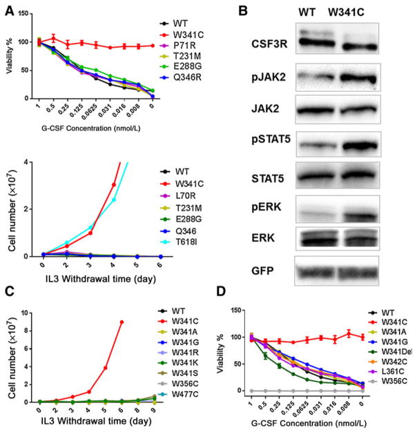
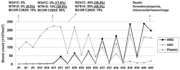
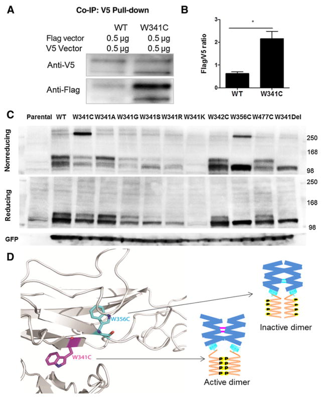
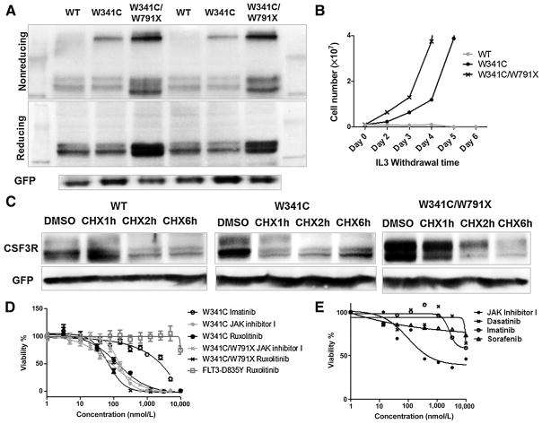
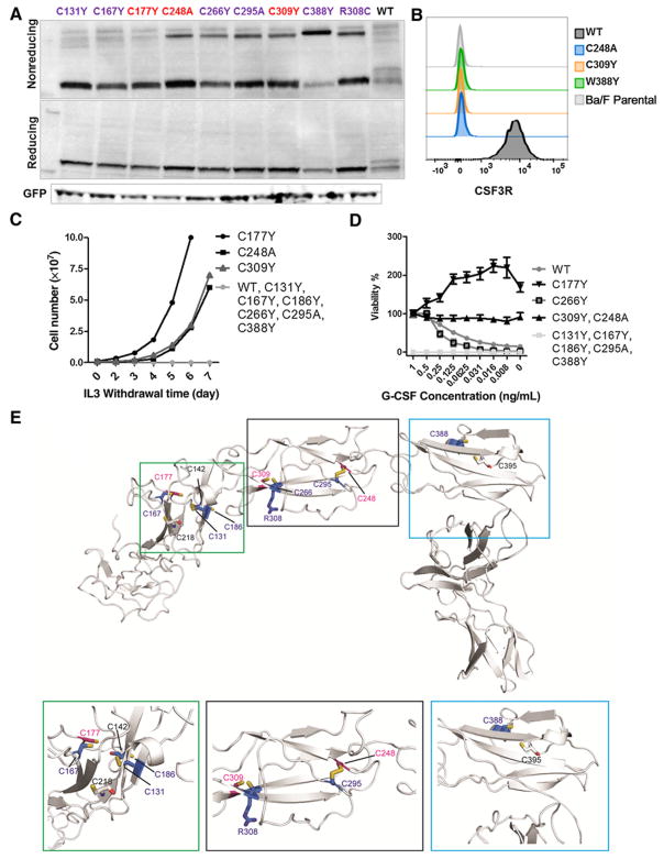
Exclusive of membrane-proximal mutations seen commonly in chronic neutrophilic leukemia (e.g., T618I), functionally defective mutations in the extracellular domain of the G-CSF receptor (CSF3R) have been reported only in severe congenital and idiopathic neutropenia patients. Here, we describe the first activating mutation in the fibronectin-like type III domain of the extracellular region of CSF3R (W341C) in a leukemia patient. This mutation transformed cells via cysteine-mediated intermolecular disulfide bonds, leading to receptor dimerization. Interestingly, a CSF3R cytoplasmic truncation mutation (W791X) found on the same allele as the extracellular mutation and the expansion of the compound mutation was associated with increased leukocytosis and disease progression of the patient. Notably, the primary patient sample and cells transformed by W341C and W341C/W791X exhibited sensitivity to JAK inhibitors. We further showed that disruption of original cysteine pairs in the CSF3R extracellular domain resulted in either gain- or loss-of-function changes, part of which was attributable to cysteine-mediated dimer formation. This, therefore, represents the first characterization of unpaired cysteines that mediate both gain- and loss-of-function phenotypes. Overall, our results show the structural and functional importance of conserved extracellular cysteine pairs in CSF3R and suggest the necessity for broader screening of CSF3R extracellular domain in leukemia patients. Cancer Res; 77(16); 4258-67. ©2017 AACR.


Conflict of interest statement

Disclosure of Potential Conflicts of Interest

J.W. Tyner reports receiving commercial research grants from Agios, Aptose, Array, AstraZeneca, Constellation, Genentech, Incyte, Janssen, Seattle Genetics, Syros, and Takeda and is a consultant/advisory board member for Leap Oncology. No potential conflicts of interest were disclosed by the other authors.

Figures

Figure 1
Figure 1. Identification of a CSF3R extracellular autoactivating mutation W341C
A, Top, Ba/F3 cells expressing CSF3R WT and mutants were plated with a concentration gradient of G-CSF after removing IL3 and cultured for 72 hours and subjected to MTS assay. Representative graph depicts percentage of cell viability (mean ± SEM) normalized to the highest G-CSF concentration (1 ng/mL) for each cell line. Bottom, representative IL3 withdrawal assay of Ba/F3 cells expressing CSF3R WT and extracellular mutations. T618I cells were used as a positive control. B, HEK293T/17 cells were transfected with CSF3R WT and W341C constructs for 48 hours. Transfected cells were then serum starved for 4 hours and subjected to immunoblot probing for CSF3R, pJAK2, pSTAT5, and pERK. C, Representative transforming assay of Ba/F3 cells expressing tryptophan to cysteine substitution at different amino acid positions and tryptophan to other amino acid substitutions at 341. D, Ba/F3 cells expressing CSF3R WT and mutants were plated with a concentration gradient of G-CSF after removing IL3 and cultured for 72 hours and subjected to MTS assay. Representative graph depicts percentage of cell viabilities (mean ± SEM) normalized to the highest G-CSF concentration (1 ng/mL) for each cell line. Images shown are representative of at least three independent experiments.
Figure 2
Figure 2
W341C/W791X causes progressively increased WBC and absolute neutrophil count of the patient. Serial WBC, platelet, and absolute neutrophil counts with mutation status during treatment. BCOR mutation was validated with Sanger sequencing. Mutations of CSF3R were validated with Sanger sequencing and targeted deep sequencing (highlighted with underline).
Figure 3
Figure 3. W341C induces intermolecular disulfide bond formation and leads to increased receptor dimerization
A, HEK293T/17 cells were transfected with equal amount of Flag- and V5-tagged CSF3R WT or W341C construct for 48 hours and subjected to co-IP assay. Representative immunoblot image demonstrates increased CSF3R pull-down in W341C cells. B, Graph depicts increased Flag/V5 ratios of CSF3R W341C compared with WT in co-IP assay of three independent experiments. Data, mean ± SEM. Statistical significance was determined using Student two-tailed t tests and expressed as P value (*, P < 0.05). C, CSF3R expression determined by nonreducing (top) and reducing immunoblots (bottom) in transfected HEK293T/17 cells expressing CSF3R variants. Protein molecular weight markers are shown on both sides. Images shown are representative of more than three independent experiments. D, Homology model of the CSF3R extracellular domain. W341 and W356 are shown in stick representation. A hypothesized model involving active and inactive dimer is illustrated.
Figure 4
Figure 4
The CSF3R W341C/W791X compound mutation demonstrates enhanced oncogenic potential. A, Representative immunoblot image of CSF3R expression of WT, W341C, and W341C/W791X compound mutation in nonreducing and reducing conditions. Protein molecular weight marker was shown on both sides. B, Representative IL3 withdrawal assay demonstrated faster outgrowth kinetics of Ba/F3 cells expressing the W341C/W791X compound mutation compared with W341C alone. C, Representative immunoblot images showing CSF3R expression in HEK293T/17 cells transiently transfected with WT, W341C, or W341C/W791X after cycloheximide (CHX; 100 mg/mL) treatment. D, Transduced Ba/F3 cells were treated with gradient concentrations of different inhibitors for 72 hours, and cell viability was assessed by MTS assay as described in Materials and Methods. Graph depicts percentages of cell viability (mean ± SEM) at different drug concentrations normalized to nondrug treatment control. Images shown are representative of at least three independent experiments. FLT3-D835Y–transformed Ba/F3 cells were used as control. E, Primary patient PBMCs were plated with graded concentrations of a panel of small-molecule inhibitors for 72 hours, and cell viability was determined by MTS assay. Graph depicts percentages of cell viability of different inhibitors at different drug concentrations normalized to nondrug treatment control.
Figure 5
Figure 5
Disruption of the intramolecular cysteine pairs demonstrates either gain- or loss-of-function phenotypes. A, CSF3R expression determined by nonreducing (top) and reducing (bottom) immunoblots in transfected HEK293T/17 cells. Protein molecular weight marker is shown on the left side. Images shown are representative of three independent experiments. B, Representative histogram depicts surface CSF3R expression. C, Representative IL3 withdrawal assay of Ba/F3 cells expressing mutations disrupting the cysteine pairs at the CSF3R extracellular domain. D, Ba/F3 cells expressing CSF3R WT and mutants were plated with a concentration gradient of G-CSF after removing IL3 and cultured for 72 hours and subjected to MTS assay. E, Homology models the CSF3R extracellular domain. The structural location of the mutants of extracellular cysteine pairs is shown in stick representation and is colored according to their phenotype after substitution of cysteine (violet, loss of function; red, gain of function; black, uncharacterized).

Similar articles

Cited by

References

    1. Maxson JE, Gotlib J, Pollyea DA, Fleischman AG, Agarwal A, Eide CA, et al. Oncogenic CSF3R mutations in chronic neutrophilic leukemia and atypical CML. N Engl J Med. 2013;368:1781–90. - PMC - PubMed
    1. Maxson JE, Luty SB, MacManiman JD, Paik JC, Gotlib J, Greenberg P, et al. The Colony Stimulating Factor 3 Receptor T640N mutation is oncogenic, sensitive to JAK inhibition, and mimics T618I. Clin Cancer Res. 2015;22:757–64. - PMC - PubMed
    1. Maxson JE, Luty SB, MacManiman JD, Abel ML, Druker BJ, Tyner JW. Ligand independence of the T618I mutation in the colony-stimulating factor 3 receptor (CSF3R) protein results from loss of O-linked glyco-sylation and increased receptor dimerization. J Biol Chem. 2014;289:5820–7. - PMC - PubMed
    1. Fleischman AG, Maxson JE, Luty SB, Agarwal A, Royer LR, Abel ML, et al. The CSF3R T618I mutation causes a lethal neutrophilic neoplasia in Mice that is responsive to therapeutic JAK inhibition. Blood. 2013;122:3628–31. - PMC - PubMed
    1. Aarts LHJ, Roovers O, Ward AC, Touw IP. Receptor activation and 2 distinct COOH-terminal motifs control G-CSF receptor distribution and internalization kinetics. Blood. 2004;103:571–9. - PubMed

Publication types