. 2017 Aug 15;77(16):4460-4471.
doi: 10.1158/0008-5472.CAN-17-0015. Epub 2017 Jun 26.

Posttranscriptional Upregulation of IDH1 by HuR Establishes a Powerful Survival Phenotype in Pancreatic Cancer Cells

Affiliations

Posttranscriptional Upregulation of IDH1 by HuR Establishes a Powerful Survival Phenotype in Pancreatic Cancer Cells

Mahsa Zarei et al. Cancer Res. .

Abstract

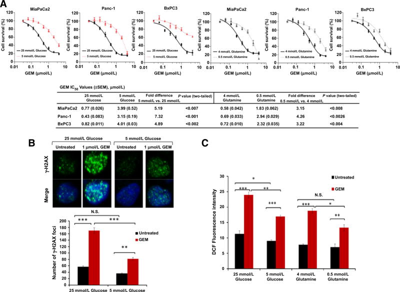
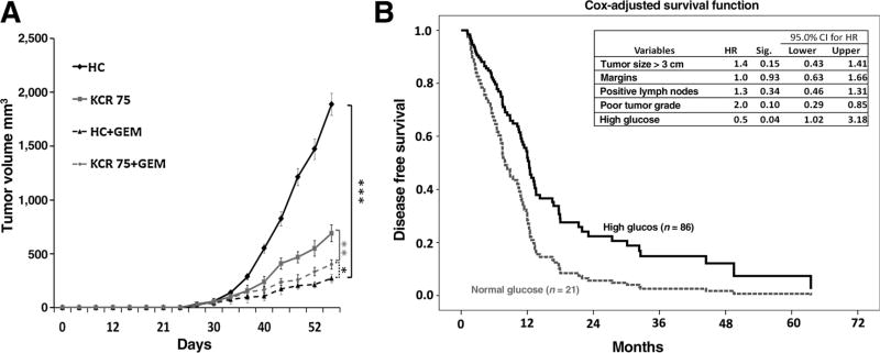
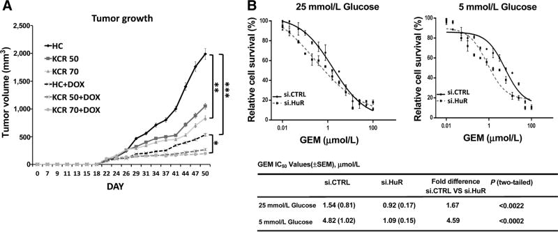
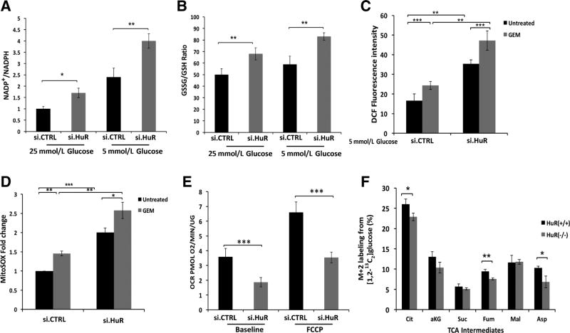
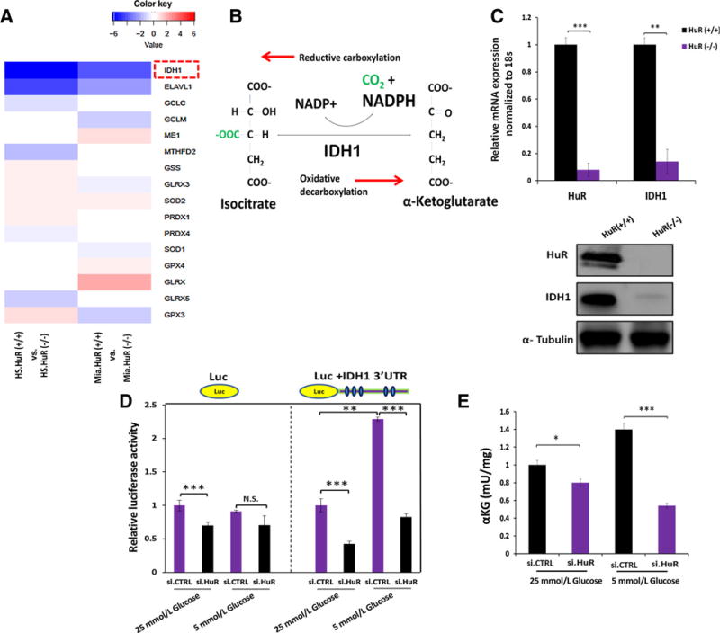
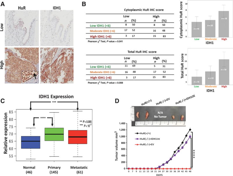
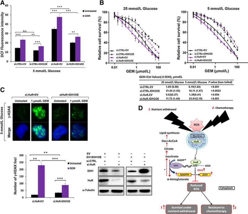
Cancer aggressiveness may result from the selective pressure of a harsh nutrient-deprived microenvironment. Here we illustrate how such conditions promote chemotherapy resistance in pancreatic ductal adenocarcinoma (PDAC). Glucose or glutamine withdrawal resulted in a 5- to 10-fold protective effect with chemotherapy treatment. PDAC xenografts were less sensitive to gemcitabine in hypoglycemic mice compared with hyperglycemic mice. Consistent with this observation, patients receiving adjuvant gemcitabine (n = 107) with elevated serum glucose levels (HgbA1C > 6.5%) exhibited improved survival. We identified enhanced antioxidant defense as a driver of chemoresistance in this setting. ROS levels were doubled in vitro by either nutrient withdrawal or gemcitabine treatment, but depriving PDAC cells of nutrients before gemcitabine treatment attenuated this effect. Mechanistic investigations based on RNAi or CRISPR approaches implicated the RNA binding protein HuR in preserving survival under nutrient withdrawal, with or without gemcitabine. Notably, RNA deep sequencing and functional analyses in HuR-deficient PDAC cell lines identified isocitrate dehydrogenase 1 (IDH1) as the sole antioxidant enzyme under HuR regulation. HuR-deficient PDAC cells lacked the ability to engraft successfully in immunocompromised mice, but IDH1 overexpression in these cells was sufficient to fully restore chemoresistance under low nutrient conditions. Overall, our findings highlight the HuR-IDH1 regulatory axis as a critical, actionable therapeutic target in pancreatic cancer. Cancer Res; 77(16); 4460-71. ©2017 AACR.


Conflict of interest statement

Disclosure of Potential Conflicts of Interest

C.M. Metallo is a consultant/advisory board member of Agios Pharmaceuticals. No potential conflicts of interest were disclosed by the other authors.

Figures

Figure 1
Figure 1. Low nutrient levels promote chemotherapy resistance in PDAC (in vitro model)
A, Survival of PDAC cell lines treated with the indicated doses of gemcitabine (GEM). IC50 values are provided. Cell survival was calculated by measurement of dsDNA content using PicoGreen. B, γ-H2AX foci. MiaPaCa2 cells cultured for 48 hours and treated with gemcitabine for the final 18 hours. C, ROS levels in MiaPaCa2 cells by DCF fluorescence and under the indicated conditions for 48 hours. Cells were treated with gemcitabine (1 µmol/L) for the final 18 hours. Error bars represent ± SEM of triplicate wells from a representative experiment. (N.S., nonsignificant; *, P < 0.05; **, P < 0.01; ***, P < 0.001). See also Supplementary Fig. S1.
Figure 2
Figure 2. Low nutrient conditions promote chemotherapy resistance in PDAC (in vivo mouse and in patients)
A, Tumor growth curves of MiaPaCa2 PDAC xenografts in mice fed an HC or a ketogenic and calorie restricted diet (75% of the average caloric intake, KCR75). Each data point represents the mean ± SEM (n= 5 per group). *, P < 0.05; **, P < 0.01; ***, P < 0.001. B, Cox multivariate disease-free survival in patients receiving gemcitabine after resection for PDAC. See also Supplementary Fig. S2. GEM, gemcitabine.
Figure 3
Figure 3. HuR protects PDAC cells against nutrient withdrawal and chemotherapy
A, Tumor growth curves of MiaPaCa2 PDAC xenografts containing a doxycycline (DOX)-inducible shRNA construct against HuR (MiaPaCa2.sh.HuR). Mice were fed an HC diet, a ketogenic diet with 75% caloric intake (KCR75), or a ketogenic diet with 50% caloric intake (KCR50). Each data point represents the mean ± SEM (n= 8 per group). B, PicoGreen cell survival and drug sensitivity assays in MiaPaCa2 cells treated with gemcitabine (GEM). See also Supplementary Figs. S3 and S4.
Figure 4
Figure 4. HuR minimizes ROS levels, while enhancing mitochondrial function
A, NADP+/NADPH ratio in MiaPaCa2 cells cultured under the indicated conditions for 24 hours. B, GSSG/GSH ratio in MiaPaCa2 cells under the same conditions. C, ROS levels, detected by DCF fluorescence in MiaPaCa2 cells cultured in 5 mmol/L glucose media for 24 hours, and gemcitabine (GEM; 1 µmol/L) as indicated. D, Mitochondrial-specific ROS were analyzed using MitoSOX in MiaPaCa2 cells cultured in 5 mmol/L glucose media for 24 hours, and gemcitabine (1 µmol/L) as indicated. E, Oxygen consumption rates (OCR) in MiaPaCa2 cells, cultured in 5 mmol/L glucose for 24 hours. F, Quantitative 13C-isotope tracer labeling of TCA cycle intermediates (or surrogate metabolites, e.g., Asp) in MiaPaca2 cells that were modified by CRISPR/Cas9 against HuR(−/−), or isogenic controls (+/+). Cells were cultured in 5 mmol/L glucose for 24 hours in the presence of [1,2-13C6]glucose. Each data point represents the mean ± SEM of three independent experiments. *, P < 0.05; **, P < 0.01; ***, P < 0.001. See also Supplementary Fig. S5.
Figure 5
Figure 5. HuR regulates IDH1 expression in PDAC
A, RNA sequencing was performed in two PDAC cell lines (HS-766T and MiaPaCa2) modulated by CRISPR/Cas9 against HuR. The heatmap displays transcripts encoding antioxidant enzymes (out of 40 directly involved in ROS detoxification), with significant changes in HuR(−/−) cells as compared with the HuR(+/+) control cells. HuR (ELAVL1, the CRISPR deletion target) is included in the table as reference. B, Schematic of the reversible IDH1 catalytic reaction. NADPH is produced by oxidative decarboxylation. C, HuR and IDH1 mRNA levels in MiaPaca2 CRISPR HuR(+/+) or HuR(−/−) cells; immunoblot analysis of IDH1 and HuR protein in the same cells. D, MiaPaCa2 cells were cotransfected with siRNAs and luciferase reporter constructs (luciferase control or fused with IDH1 3′ UTR). Cells were cultured as indicated for 24 hours. E, αKG levels in MiaPaCa2 cells cultured under the indicated conditions for 24 hours. Error bars, ± SEM of triplicate wells from a representative experiment. *, P < 0.05; **, P < 0.01; ***, P < 0.001. See also Supplementary Figs. S6 and S7.
Figure 6
Figure 6. HuR is associated with IDH1 expression in clinical PDAC samples
A, Immunohistochemistry of HuR and IDH1 in formalin-fixed, paraffin-embedded PDAC tumors. The inset and arrow indicate cytoplasmic HuR expression. B, Tabulation and graphing of IHC scores from the TMA dataset shows cytoplasmic and total HuR IHC scores to be associated with IDH1 scores. Spearman correlation test yielded a significant correlation between the scores (P = 0.33, P < 0.01). Error bars display 95% confidence interval. C, IDH1 mRNA expression levels in normal pancreatic tissue, primary PDAC, and metastatic PDAC. For each boxplot, median expression value, and first and third quartiles are indicated. ***, P < 0.005; ****, P < 0.0000001. D, Representative images of subcutaneous MiaPaca2 xenografts (day 48) along with tumor growth curves. Each data point represents the mean ± SEM (n= 8 per group; ****, P < 0.0001). See also Supplementary Fig. S8.
Figure 7
Figure 7. IDH1 rescues HuR-deficient PDAC cells from metabolic stress
A, ROS levels in MiaPaCa2 as measured by DCF fluorescence. The indicated culture conditions lasted 48 hours, with gemcitabine (GEM; 1 µmol/L) treatment for the final 18 hours, as indicated. Cotransfections with siRNAs and overexpression plasmids were performed as indicated and repeated for B and C. B, PicoGreen cell survival and drug sensitivity assays in MiaPaCa2 cells. C, γ-H2AX foci in MiaPaCa2 cells. Culture and treatments paralleled the experiment in A. D, A schematic model of how HuR’s regulation of IDH1 promotes survival under nutrient withdrawal and chemotherapy treatment by enhancing reductive power and antioxidant defense. Error bars, ± SEM of triplicate wells from a representative experiment (N.S., nonsignificant; **, P < 0.01; ***, P < 0.001). See also Supplementary Fig. S8.

Similar articles

Cited by

References

    1. Kamphorst JJ, Nofal M, Commisso C, Hackett SR, Lu W, Grabocka E, et al. Human pancreatic cancer tumors are nutrient poor and tumor cells actively scavenge extracellular protein. Cancer Res. 2015;75:544–53. - PMC - PubMed
    1. Provenzano PP, Cuevas C, Chang AE, Goel VK, Von Hoff DD, Hingorani SR. Enzymatic targeting of the stroma ablates physical barriers to treatment of pancreatic ductal adenocarcinoma. Cancer Cell. 2012;21:418–29. - PMC - PubMed
    1. Ros PR, Mortele KJ. Imaging features of pancreatic neoplasms. JBR-BTR. 2001;84:239–49. - PubMed
    1. Morgan G, Ward R, Barton M. The contribution of cytotoxic chemotherapy to 5-year survival in adult malignancies. Clin Oncol. 2004;16:549–60. - PubMed
    1. Conroy T, Desseigne F, Ychou M, Bouche O, Guimbaud R, Becouarn Y, et al. FOLFIRINOX versus gemcitabine for metastatic pancreatic cancer. N Engl J Med. 2011;364:1817–25. - PubMed

MeSH terms