. 2017 Sep 5;199(19):e00162-17.
doi: 10.1128/JB.00162-17. Print 2017 Oct 1.

Metabolism Dealing with Thermal Degradation of NAD+ in the Hyperthermophilic Archaeon Thermococcus kodakarensis

Affiliations

Metabolism Dealing with Thermal Degradation of NAD+ in the Hyperthermophilic Archaeon Thermococcus kodakarensis

Shin-Ichi Hachisuka et al. J Bacteriol. .

Abstract

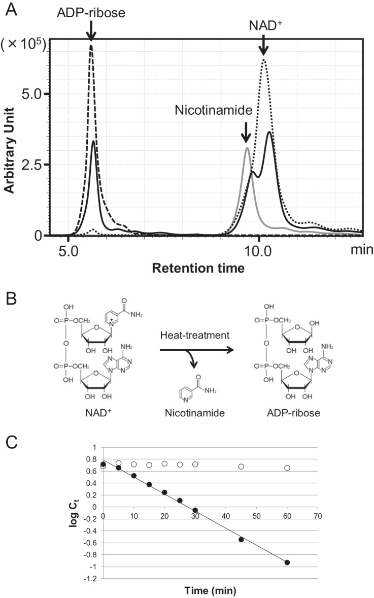
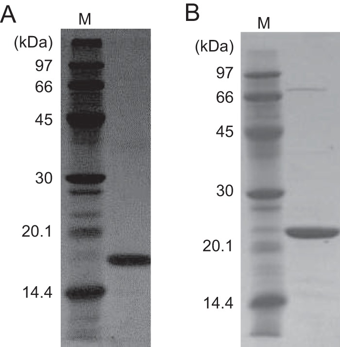
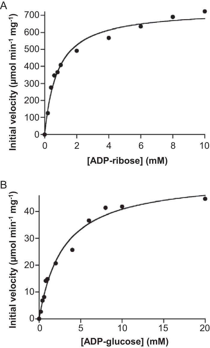
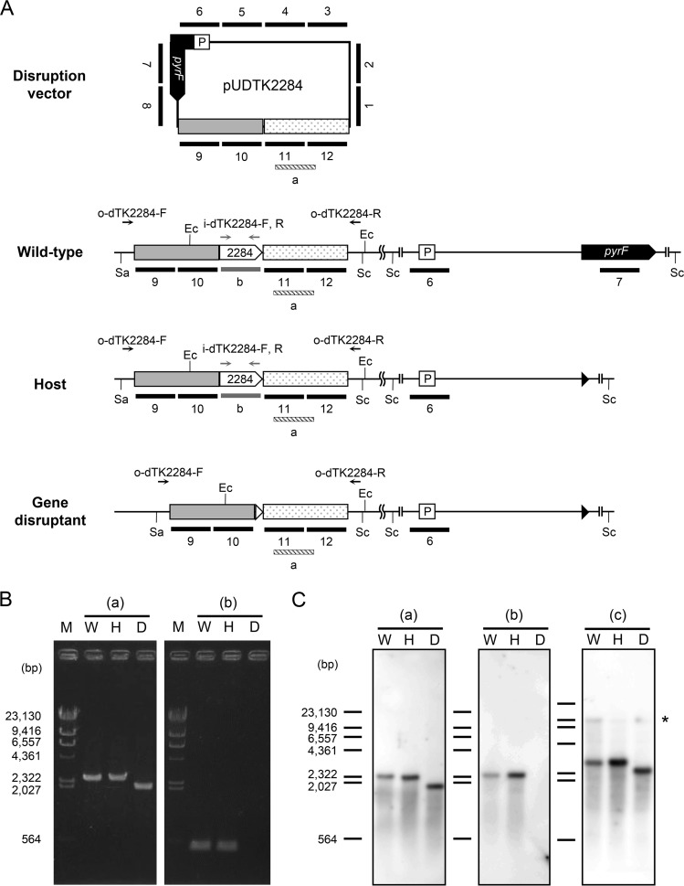
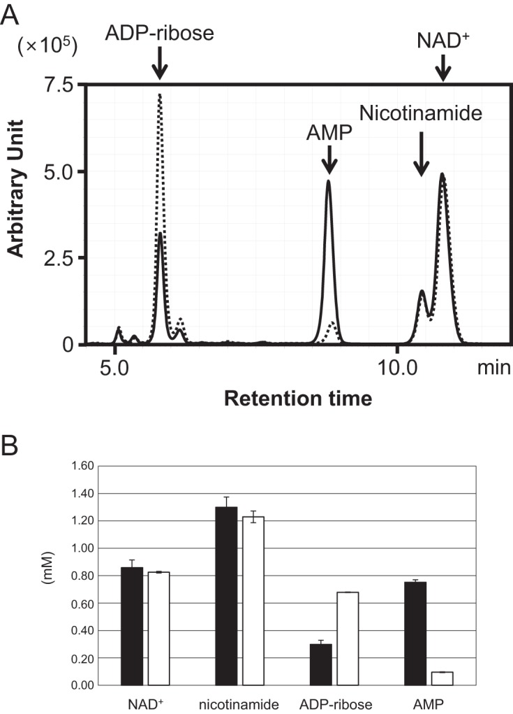
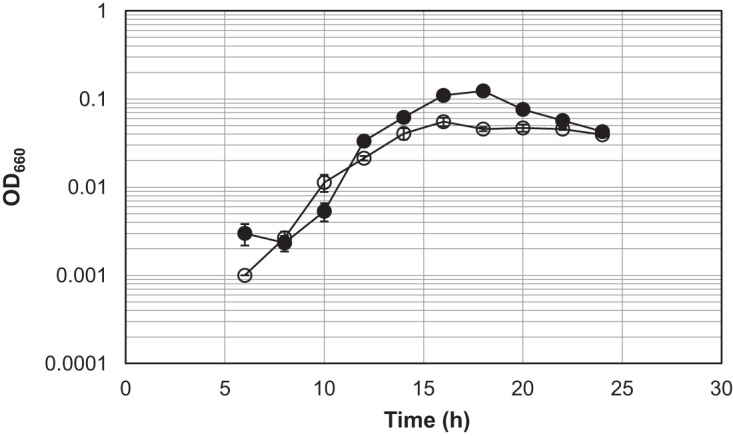
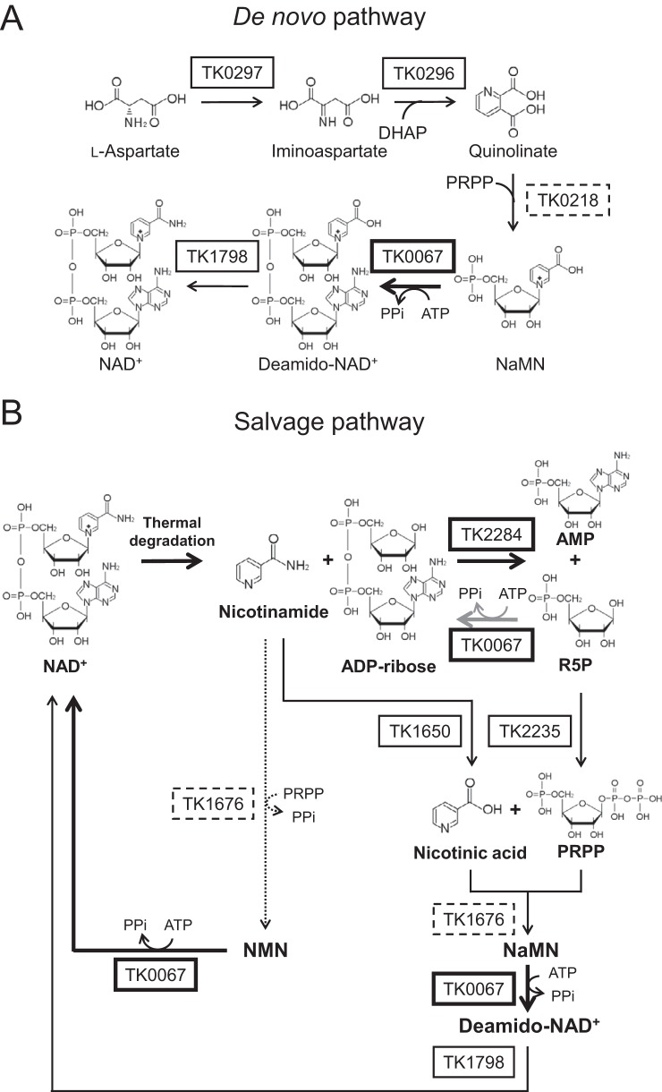
NAD+ is an important cofactor for enzymatic oxidation reactions in all living organisms, including (hyper)thermophiles. However, NAD+ is susceptible to thermal degradation at high temperatures. It can thus be expected that (hyper)thermophiles harbor mechanisms that maintain in vivo NAD+ concentrations and possibly remove and/or reuse undesirable degradation products of NAD+ Here we confirmed that at 85°C, thermal degradation of NAD+ results mostly in the generation of nicotinamide and ADP-ribose, the latter known to display toxicity by spontaneously linking to proteins. The hyperthermophilic archaeon Thermococcus kodakarensis possesses a putative ADP-ribose pyrophosphatase (ADPR-PPase) encoded by the TK2284 gene. ADPR-PPase hydrolyzes ADP-ribose to ribose 5-phosphate (R5P) and AMP. The purified recombinant TK2284 protein exhibited activity toward ADP-ribose as well as ADP-glucose. Kinetic analyses revealed a much higher catalytic efficiency toward ADP-ribose, suggesting that ADP-ribose was the physiological substrate. To gain insight into the physiological function of TK2284, a TK2284 gene disruption strain was constructed and examined. Incubation of NAD+ in the cell extract of the mutant strain at 85°C resulted in higher ADP-ribose accumulation and lower AMP production compared with those in experiments with the host strain cell extract. The mutant strain also exhibited lower cell yield and specific growth rates in a synthetic amino acid medium compared with those of the host strain. The results obtained here suggest that the ADPR-PPase in T. kodakarensis is responsible for the cleavage of ADP-ribose to R5P and AMP, providing a means to utilize the otherwise dead-end product of NAD+ breakdown.IMPORTANCE Hyperthermophilic microorganisms living under high temperature conditions should have mechanisms that deal with the degradation of thermolabile molecules. NAD+ is an important cofactor for enzymatic oxidation reactions and is susceptible to thermal degradation to ADP-ribose and nicotinamide. Here we show that an ADP-ribose pyrophosphatase homolog from the hyperthermophilic archaeon Thermococcus kodakarensis converts the detrimental ADP-ribose to ribose 5-phosphate and AMP, compounds that can be directed to central carbon metabolism. This physiological role for ADP-ribose pyrophosphatases might be universal in hyperthermophiles, as their homologs are widely distributed among both hyperthermophilic bacteria and archaea.

Keywords: ADP-ribose pyrophosphatase; Archaea; NAD+; Thermococcus; hyperthermophiles; thermal degradation.


Figures

FIG 1
FIG 1
Identification of the thermal degradation products of NAD+ and determination of the thermal degradation rates of NAD+. (A) HPLC analysis was performed to identify the thermal degradation products of NAD+. The solid line shows NAD+ after heat treatment at 85°C for 30 min. The dotted line shows an NAD+ standard, the dashed line shows an ADP-ribose standard, and the gray line shows a nicotinamide standard. All samples were analyzed by monitoring A215. (B) Chemical reaction of the thermal degradation of NAD+. (C) Thermal degradation rate of NAD+ and ADP-ribose at 85°C. Ct indicates concentration of NAD+ or ADP-ribose (millimolar) after heat treatment for t min. log Ct indicates natural logarithm of Ct. Filled circles and open circles indicate NAD+ and ADP-ribose, respectively.
FIG 2
FIG 2
Purified recombinant TK2284 and TK0067 proteins. Purified TK2284 (A) and TK0067 (B) recombinant proteins were analyzed by SDS-PAGE and stained with Coomassie brilliant blue. Four micrograms of protein was applied in each lane.
FIG 3
FIG 3
Kinetic analyses of the hydrolase reactions toward ADP-ribose (A) and ADP-glucose (B). Initial velocities were measured in the presence of various concentrations of ADP-ribose and ADP-glucose.
FIG 4
FIG 4
Genotypic analyses of the ADPR-PPase gene deletion mutant. (A) Schematic drawing of relevant regions of the pUDTK2284 vector and the chromosome in wild-type (KOD1), host (KU216), and the TK2284 disruption strains. Primer sets used in PCR analyses, annealing within the TK2284 coding region (i-dTK2284-F/i-dTK2284-R) and outside the homologous regions (o-dTK2284-F/o-dTK2284-R), are indicated with gray and black arrows, respectively. Probes annealing to regions within the TK2284 gene, the 3′-flanking region of the TK2284 gene, and the entire sequence of pUDTK2284 in Southern blot analysis are indicated with gray, striped, and black bars, respectively. Although there are gaps between bars 1 to 12, the probes actually cover the entire sequence of pUDTK2284 without any gaps. The cleavage sites of the restriction enzymes used in Southern blot analyses are also shown. (B) PCR analyses of the TK2284 gene deletion mutant were performed with primer sets that anneal outside the homologous regions for homologous recombination (a) and within the TK2284 gene (b). (C) Southern blot analyses were performed with probe a binding to the 3′-flanking region of the TK2284 gene (a), probe b binding within the gene (b), and probes 1 to 12 corresponding to the entire sequence of the pUDTK2284 vector (c). The asterisk indicates the signals corresponding to fragments containing the pyrF promoter and/or pyrF gene (9.8 kbp in the wild-type strain and 9.2 kbp in the host strain and the deletion mutant). Ec, cleavage site of EcoRI; Sa, cleavage site of SalI; Sc, cleavage site of ScaI; P, promoter region of the operon including pyrF; M, marker; W, T. kodakarensis wild-type KOD1; H, T. kodakarensis host strain KU216; D, TK2284 gene disruptant.
FIG 5
FIG 5
Thermal degradation of NAD+ and enzymatic degradation of ADP-ribose in cell extracts. (A) NAD+ (2 mM) and MgCl2 (10 mM) were added to cell extracts and heat treated at 85°C for 30 min. Solid and dotted lines show products in cell extracts from the KU216 host strain and TK2284 gene disruptant, respectively. Both samples were analyzed by monitoring A254. The experiment was repeated with cell extracts obtained from three independent cultures for each strain, and a representative result is shown. (B) The concentrations of NAD+, nicotinamide, ADP-ribose, and AMP were calculated with the results shown in panel A, and the averages and standard deviations (n = 3) are shown. Filled bars indicate results with the cell extract of the KU216 host strain, and open bars indicate those of the gene disruptant.
FIG 6
FIG 6
Growth properties of the T. kodakarensis host strain KU216 and TK2284 gene disruptant. Both strains were grown in a synthetic amino acid medium, ASW-AA-S0-Pyr-Ura-W. Filled circles and open circles represent the results from three independent growth experiments with the host strain and the TK2284 gene disruptant, respectively. The vertical axis is represented in logarithmic scale.
FIG 7
FIG 7
Predicted NAD+ biosynthesis and salvage pathways in T. kodakarensis. (A) De novo NAD+ biosynthesis pathway from aspartate. (B) Salvage pathway for NAD+ regeneration predicted from genomic information and the results obtained in this study and elsewhere (34–38). Bold arrows represent reactions examined in this study and the gray arrow indicates the side reaction of the TK0067 protein. Solid arrows show generally predicted pathways in Archaea and the dotted arrow indicates the reaction proposed in Discussion. Proteins examined in this study are in bold boxes, proteins that have been examined in other archaea are in solid boxes, and proteins only predicted by amino acid sequence and/or structure are in dashed boxes. DHAP, dihydroxyacetone phosphate; deamido-NAD+, nicotinic acid adenine dinucleotide; PRPP, phosphoribosyl pyrophosphate.

Similar articles

Cited by

References

    1. Dwivedi N, Dube D, Pandey J, Singh B, Kukshal V, Ramachandran R, Tripathi RP. 2008. NAD+-dependent DNA ligase: a novel target waiting for the right inhibitor. Med Res Rev 28:545–568. doi:10.1002/med.20114. - DOI - PubMed
    1. Hassa PO, Haenni SS, Elser M, Hottiger MO. 2006. Nuclear ADP-ribosylation reactions in mammalian cells: where are we today and where are we going? Microbiol Mol Biol Rev 70:789–829. doi:10.1128/MMBR.00040-05. - DOI - PMC - PubMed
    1. Hassa PO, Hottiger MO. 2008. The diverse biological roles of mammalian PARPS, a small but powerful family of poly-ADP-ribose polymerases. Front Biosci 13:3046–3082. doi:10.2741/2909. - DOI - PubMed
    1. Song SH, Ahluwalia N, Leduc Y, Delbaere LT, Vieille C. 2008. Thermotoga maritima TM0298 is a highly thermostable mannitol dehydrogenase. Appl Microbiol Biotechnol 81:485–495. doi:10.1007/s00253-008-1633-9. - DOI - PubMed
    1. Kallnik V, Schulz C, Schweiger P, Deppenmeier U. 2011. Properties of recombinant Strep-tagged and untagged hyperthermophilic d-arabitol dehydrogenase from Thermotoga maritima. Appl Microbiol Biotechnol 90:1285–1293. doi:10.1007/s00253-011-3187-5. - DOI - PubMed

Publication types

LinkOut - more resources