. 2017 Jul 11;114(28):7367-7372.
doi: 10.1073/pnas.1707386114. Epub 2017 Jun 26.

Cryo-EM structure of the DNA-PK holoenzyme

Affiliations

Cryo-EM structure of the DNA-PK holoenzyme

Humayun Sharif et al. Proc Natl Acad Sci U S A. .

Abstract

DNA-dependent protein kinase (DNA-PK) is a large protein complex central to the nonhomologous end joining (NHEJ) DNA-repair pathway. It comprises the DNA-PK catalytic subunit (DNA-PKcs) and the heterodimer of DNA-binding proteins Ku70 and Ku80. Here, we report the cryo-electron microscopy (cryo-EM) structures of human DNA-PKcs at 4.4-Å resolution and the DNA-PK holoenzyme at 5.8-Å resolution. The DNA-PKcs structure contains three distinct segments: the N-terminal region with an arm and a bridge, the circular cradle, and the head that includes the kinase domain. Two perpendicular apertures exist in the structure, which are sufficiently large for the passage of dsDNA. The DNA-PK holoenzyme cryo-EM map reveals density for the C-terminal globular domain of Ku80 that interacts with the arm of DNA-PKcs. The Ku80-binding site is adjacent to the previously identified density for the DNA-binding region of the Ku70/Ku80 complex, suggesting concerted DNA interaction by DNA-PKcs and the Ku complex.

Keywords: DNA repair; NHEJ; V(D)J recombination; cryo-EM; structure.


Conflict of interest statement

The authors declare no conflict of interest.

Figures
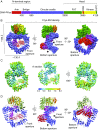
Fig. 1.
Fig. 1.
The overall DNA-PKcs structure. (A) Schematics of DNA-PKcs domain architecture showing the three units of the DNA-PKcs structure: N-terminal region with arm (red) and bridge (magenta), the circular cradle (blue) and the head domain containing FAT (FRAP-ATM-TRRAP) (green), FRB (FKBP12-rapamycin-binding) (orange), kinase (yellow), and FATC (FAT C-terminal) (dark green) domains. (B) Cryo-EM density of DNA-PKcs at 4.4-Å resolution. (C) Overall resolution distribution of DNA-PKcs cryo-EM map. Two different views are shown on the Left and Right. (Middle) Cut-through section of DNA-PKcs. The resolution range is color-coded. (D) Cartoon representation of DNA-PKcs model in orientations similar to in B.
Fig. S1.
Fig. S1.
The overall DNA-PKcs structure. (A) SDS/PAGE of two fractions of the DNA-PK sample eluted from the last Superose 6 step of the purification. The right lane was used for cryo-EM data collection. (B) A cryo-EM micrograph of DNA-PK (Upper) and five 2D classes of DNA-PK (Bottom). (C) Angular distribution for the initial DNA-PK data set (∼860K particles) (Left) and filtered data set (∼290K particles) that removed two-thirds of particles at the preferred orientation (Right). Each cylinder represents one view, and the height is scaled to the number of particles in that view. (D) The FSC curve of the final refined cryo-EM map of DNA-PKcs at an estimated resolution of 4.4 Å.
Fig. S2.
Fig. S2.
Segments of DNA-PKcs represented in sticks fitted in the 4.4-Å-resolution cryo-EM map.
Fig. S3.
Fig. S3.
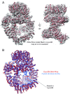
(A) DNA-PKcs model fitted in the cryo-EM density map (B) Superposition of DNA-PKcs models from cryo-EM (red) and from the crystal structure (PDB: 5LUQ) (blue). The segment that was absent in the cryo-EM map but was built as unassigned sequences in the crystal structure is indicated.
Fig. 2.
Fig. 2.
Conformational dynamics of the DNA-PKcs arm. Variations in the arm conformation in six classes of cryo-EM data are shown as cartoons: class 1 (cyan), class 2 (teal), class 3 (wheat), class 4 (light green), class 5 (light magenta), and class 6 (pink).
Fig. 3.
Fig. 3.
DNA-PK cryo-EM structure reveals Ku80 CTR binding site. (A) Schematics of Ku70 and Ku80 domains. (B) Cryo-EM density of refined DNA-PK in class 2 at 5.8-Å resolution. (Left) Two panels showing the proposed Ku80 CTR density (cyan). (Right) Two panels showing the resolution distribution of DNA-PK with color-coded resolution range. (C) DNA-PK structure with highlighted structural features: arm (red), Ku80 CTR globular domain (cyan), and modeled Ku80 CTR linker and last helix fragments from the crystal structure [Protein Data Bank (PDB): 5LUQ] (teal). (D) Ku80 CTR globular domain (cyan) may have moved the arm (red) upward in the class 2 map. If the Ku80 CTR globular domain exists in the class 6 map, it would have been in clash with the arm (purple).
Fig. S4.
Fig. S4.
DNA-PK cryo-EM structure reveals Ku80 CTR binding site. (A) Six classes of DNA-PKcs: class 1 (yellow), class 2 (blue), class 3 (pink), class 4 (salmon), class 5 (green), and class 6 (wheat). Extra density of Ku80 CTR is shown in cyan. The percentage of the total number of particles included is indicated at the bottom of each of the classes. (B) The FSC curve of refined class2 cryo-EM map at an estimated resolution of 5.8 Å. (C) Ku80 CTR globular domain NMR structure (PDB: 1Q2Z) fitted in the segmented extra density of class 2.
Fig. 4.
Fig. 4.
A structural model for the DNA-PK interaction with DNA. (A) Two views of a previously reported DNA-PK map at 25-Å resolution (EMD-1209, gray), fitted with our DNA-PK structure (gray) that contains DNA-PKcs and Ku80 CTR, and with the Ku70/80 core crystal structure (PDB: 1JEY, pink and slate). The fitted orientation of the Ku70/80 core is only approximate and was achieved by placing the Ku80 core close to the Ku80 CTR. The circle highlights the Ku70/80 core domains that mediate DNA binding. The proposed movement of the core domains resulting from the flexible linker between the Ku80 core and the CTR is shown as dashed black curvy lines. (B) 30-bp DNA duplex (black) modeled in the DNA-PK structure. (Left) DNA distal end at the front aperture. (Right) DNA free end at the bottom aperture.
Fig. S5.
Fig. S5.
A structural model for the interaction of DNA-PK with DNA. (A) Two views of DNA-PK map at 25-Å resolution (EMD-1209, gray) fitted with the DNA-PK structure, Ku70/80 crystal structure (PDB: 1JEY, pink and slate) and modeled with Ku80 C-terminal linker and helices from PDB: 5LUQ and a 30-bp duplex DNA. (B) DNA-PK cryo-EM structure dimer modeled with DNA duplex (black), Ku70/80 (PDB: 1JEY, pink and slate) in cryo-EM density of DNA-PK (EMD-1209, gray). The dimer orientation is derived from EMD-1210.

References

    1. Khanna KK, Jackson SP. DNA double-strand breaks: Signaling, repair and the cancer connection. Nat Genet. 2001;27:247–254. - PubMed
    1. Lieber MR. The mechanism of double-strand DNA break repair by the nonhomologous DNA end-joining pathway. Annu Rev Biochem. 2010;79:181–211. - PMC - PubMed
    1. Alt FW, Zhang Y, Meng FL, Guo C, Schwer B. Mechanisms of programmed DNA lesions and genomic instability in the immune system. Cell. 2013;152:417–429. - PMC - PubMed
    1. Downs JA, Jackson SP. A means to a DNA end: The many roles of Ku. Nat Rev Mol Cell Biol. 2004;5:367–378. - PubMed
    1. Smith GC, Jackson SP. The DNA-dependent protein kinase. Genes Dev. 1999;13:916–934. - PubMed

Publication types

Associated data

LinkOut - more resources