Comparative Study
. 2017 Sep 4;216(9):2669-2677.
doi: 10.1083/jcb.201612195. Epub 2017 Jun 26.

Structural differences between yeast and mammalian microtubules revealed by cryo-EM

Affiliations
Comparative Study

Structural differences between yeast and mammalian microtubules revealed by cryo-EM

Stuart C Howes et al. J Cell Biol. .

Abstract

Microtubules are polymers of αβ-tubulin heterodimers essential for all eukaryotes. Despite sequence conservation, there are significant structural differences between microtubules assembled in vitro from mammalian or budding yeast tubulin. Yeast MTs were not observed to undergo compaction at the interdimer interface as seen for mammalian microtubules upon GTP hydrolysis. Lack of compaction might reflect slower GTP hydrolysis or a different degree of allosteric coupling in the lattice. The microtubule plus end-tracking protein Bim1 binds yeast microtubules both between αβ-tubulin heterodimers, as seen for other organisms, and within tubulin dimers, but binds mammalian tubulin only at interdimer contacts. At the concentrations used in cryo-electron microscopy, Bim1 causes the compaction of yeast microtubules and induces their rapid disassembly. Our studies demonstrate structural differences between yeast and mammalian microtubules that likely underlie their differing polymerization dynamics. These differences may reflect adaptations to the demands of different cell size or range of physiological growth temperatures.


Figures

Figure 1.
Figure 1.
Lattice distinctions for yeast MTs. Models of two tubulin dimers from a PF for the indicated state. Expanded lattices (epothilone, GMPCPP, and dynamic) and compacted lattices (Dyn+Bim1, GTPγS+Bim1, and GTPγS) are distinguished by compaction across the interdimer interface, reducing the interdimer nucleotide distance (indicated by red arrows). Distances between nucleotides are given for epothilone and GTPγS (see Table S1 for exact values). Epothilone (dark blue), GTP (purple), GDP (dark green), GMPCPP (orange), and GTPγS (red) are shown with space-filled spheres; α-tubulin is shown in green and β-tubulin in blue. Note that in the dynamic lattice, a mixture of GTP and GDP is likely present at the E-site (see main text).
Figure 2.
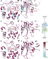
Figure 2.
Tubulin conservation around the E-site. (A and B) View from plus end (A) and minus end (B) of the MT showing sequence conservation around the E-site nucleotide between yeast and mammalian tubulin. Identical and nonidentical residues are colored maroon and cyan, respectively. Nonconservative substitutions are labeled, with mammalian residue first, whereas conservative changes are not labeled. The changes (labeled in red text) at positions 98 on β-tubulin and 253 on α-tubulin potentially create a tighter pocket for the nucleotide in yeast tubulin. (C–F) Conservation of residues surrounding the E-site across fungal tubulins (C and D) and mammalian tubulins (E and F). Fungal tubulins are generally less conserved than mammalian tubulins, though similar positions (end of H10 and position 353 in S9, both on α-tubulin) are less conserved for both.
Figure 3.
Figure 3.
Bim1 binds yeast microtubules with a monomer repeat binding pattern. (A–C) Raw images of MTs assembled from yeast (A and C) or mammalian (B) tubulin decorated with the +TIP Bim1 (A and B) or human EB3 (C). (D–F) Corresponding power spectra show the tubulin monomer repeat of 1/40 Å−1 but no +TIP repeat at 1/80 Å−1 for yeast MTs decorated with Bim1, for which Bim1 binds every tubulin monomer instead of between dimers. (G–I) Schematic (G) showing the extra, noncanonical binding position of +TIP (enlarged in H) at the intradimer contact and the canonical position (enlarged in I) adjacent to the interdimer interface. Analysis of sequence differences between yeast and mammalian proteins shows that both tubulin and +TIP sequence differences likely contribute to this binding pattern. Several significant differences are present on the lefthand side of the CH domain (+TIP residues 67–60). (J) Sequence alignment of +TIP CH domains showing significant amino acid differences involved in contacts (purple) and residues within 4 Å of tubulin (green boxes). The +TIP sequences are less conserved than tubulin, though both contribute to the binding pattern. (K) Reconstruction of yeast GTPγS MT decorated with Bim1 showing Bim1 at both inter- and intradimer sites. Docked EB3 model is shown in purple and tubulin monomers in blue (note that α- and β-tubulin cannot be distinguished in this reconstruction).

Similar articles

Cited by

References

    1. Adams P.D., Afonine P.V., Bunkóczi G., Chen V.B., Echols N., Headd J.J., Hung L.-W., Jain S., Kapral G.J., Grosse Kunstleve R.W., et al. . 2011. The Phenix software for automated determination of macromolecular structures. Methods. 55:94–106. 10.1016/j.ymeth.2011.07.005 - DOI - PMC - PubMed
    1. Akhmanova A., and Steinmetz M.O.. 2008. Tracking the ends: A dynamic protein network controls the fate of microtubule tips. Nat. Rev. Mol. Cell Biol. 9:309–322. 10.1038/nrm2369 - DOI - PubMed
    1. Alushin G.M., Ramey V.H., Pasqualato S., Ball D.A., Grigorieff N., Musacchio A., and Nogales E.. 2010. The Ndc80 kinetochore complex forms oligomeric arrays along microtubules. Nature. 467:805–810. 10.1038/nature09423 - DOI - PMC - PubMed
    1. Alushin G.M., Lander G.C., Kellogg E.H., Zhang R., Baker D., and Nogales E.. 2014. High-resolution microtubule structures reveal the structural transitions in αβ-tubulin upon GTP hydrolysis. Cell. 157:1117–1129. 10.1016/j.cell.2014.03.053 - DOI - PMC - PubMed
    1. Ayaz P., Ye X., Huddleston P., Brautigam C.A., and Rice L.M.. 2012. A TOG:αβ-tubulin complex structure reveals conformation-based mechanisms for a microtubule polymerase. Science. 337:857–860. 10.1126/science.1221698 - DOI - PMC - PubMed

Publication types

MeSH terms