

Vascular Graft Impregnation with Antibiotics: The Influence of High Concentrations of Rifampin, Vancomycin, Daptomycin, and Bacteriophage Endolysin HY-133 on Viability of Vascular Cells
- PMID: 28652563
- PMCID: PMC5498120
- DOI: 10.12659/msmbr.902879
Vascular Graft Impregnation with Antibiotics: The Influence of High Concentrations of Rifampin, Vancomycin, Daptomycin, and Bacteriophage Endolysin HY-133 on Viability of Vascular Cells
Abstract
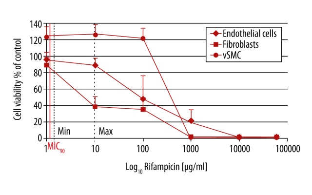
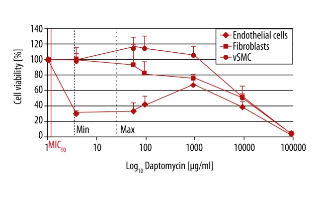
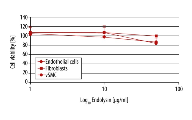
BACKGROUND Rifampin-soaked synthetic prosthetic grafts have been widely used for prevention or treatment of vascular graft infections (VGIs). This in vitro study investigated the effect of the antibiotics daptomycin and vancomycin and the new recombinant bacteriophage endolysin HY-133 on vascular cells, as potential alternatives compared to rifampin. MATERIAL AND METHODS Primary human ECs, vascular smooth muscle cells (vSMC), and fibroblasts were cultivated in 96-well plates and incubated with rifampin, daptomycin, vancomycin, and endolysin HY-133 for 24 h. Subsequently, after washing, cell viability was determined by measuring mitochondrial ATP concentration. Antibiotics were used in their corresponding minimum and maximum serum concentrations, in decimal multiples and in maximum soaking concentration. The experiments were performed in triplicate. RESULTS The 10-fold max serum concentrations of rifampin, daptomycin, and vancomycin did not influence viability of EC and vSMC (100 µg/ml, p>0.170). Higher concentrations of rifampin (>1 mg/ml) significantly (p<0.001) reduced cell viability of all cell types. For the other antibiotics, high concentrations (close to maximum soaking concentration) were most cytotoxic for EC and vSMC and fibroblasts (p<0.001). Endolysin did not display any cytotoxicity towards vascular cells. CONCLUSIONS Results of this in vitro study show the high cytotoxicity of rifampin against vascular cells, and may re-initiate the discussion about the benefit of prophylactic pre-soaking in high concentrations of rifampin. Further studies are necessary to determine the influence of rifampin on the restoration of vessel functionality versus its prophylactic effect against VGIs. Future use of recombinant phage endolysins for alternative prophylactic strategies needs further investigations.
Conflict of interest statement
None.
Figures

References
-
- Rychlik IJ, Davey P, Murphy J, O’Donnell ME. A meta-analysis to compare Dacron versus polytetrafluroethylene grafts for above-knee femoropopliteal artery bypass. J Vasc Surg. 2014;60:506–15. - PubMed
-
- Zilla P, Bezuidenhout D, Human P. Prosthetic vascular grafts: wrong models, wrong questions and no healing. Biomaterials. 2007;28:5009–27. - PubMed
-
- Dorigo W, Pulli R, Piffaretti G, et al. Results from an Italian multicentric registry comparing heparin-bonded ePTFE graft and autologous saphenous vein in below-knee femoro-popliteal bypasses. J Cardiovasc Surg. 2012;53:187–94. - PubMed
-
- Neville RF, Capone A, Amdur R, et al. A comparison of tibial artery bypass performed with heparin-bonded expanded polytetrafluoroethylene and great saphenous vein to treat critical limb ischemia. J Vasc Surg. 2012;56:1008–14. - PubMed
-
- Chaufour X, Gaudric J, Goueffic Y, et al. A multicenter experience with infected abdominal aortic endograft explantation. J Vasc Surg. 2017;65:372–80. - PubMed
MeSH terms
Substances
LinkOut - more resources
Full Text Sources
Medical
