. 2017 Jun 2:12:4195-4208.
doi: 10.2147/IJN.S132190. eCollection 2017.

Biodegradable and biocompatible cationic polymer delivering microRNA-221/222 promotes nerve regeneration after sciatic nerve crush

Affiliations

Biodegradable and biocompatible cationic polymer delivering microRNA-221/222 promotes nerve regeneration after sciatic nerve crush

Jialin Song et al. Int J Nanomedicine. .

Abstract

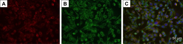
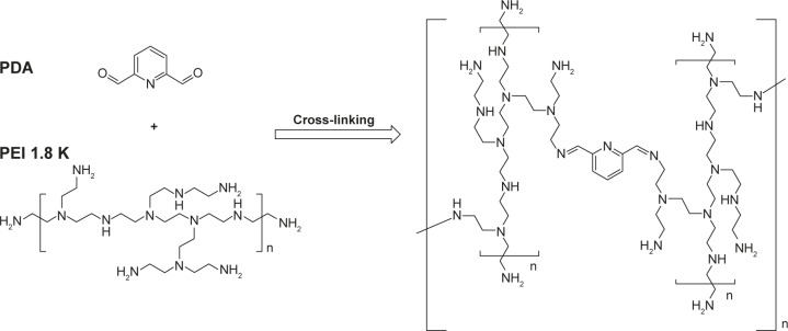
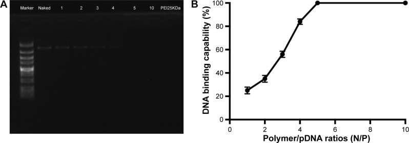
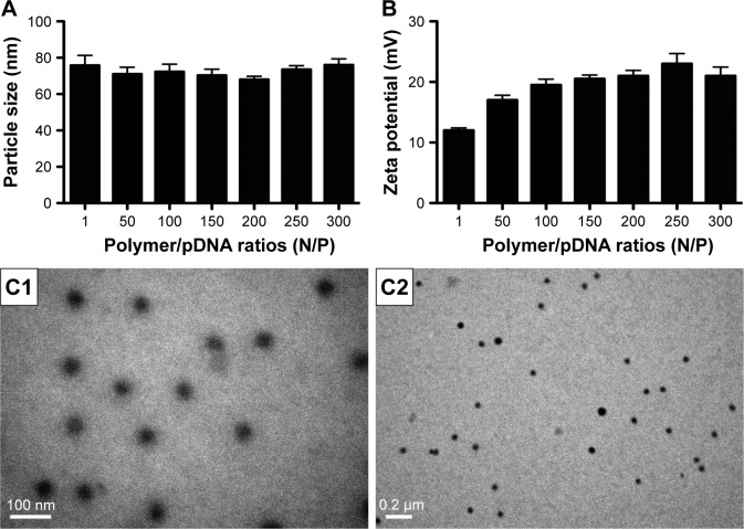
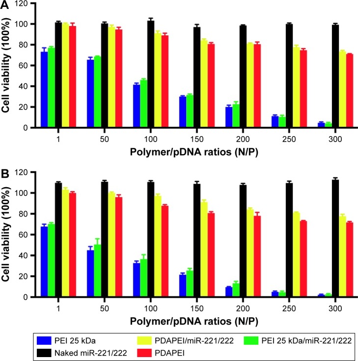
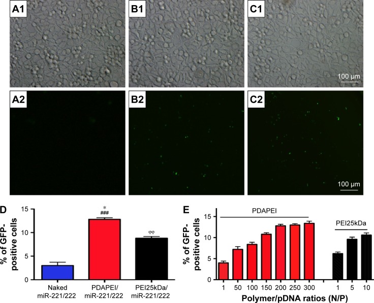
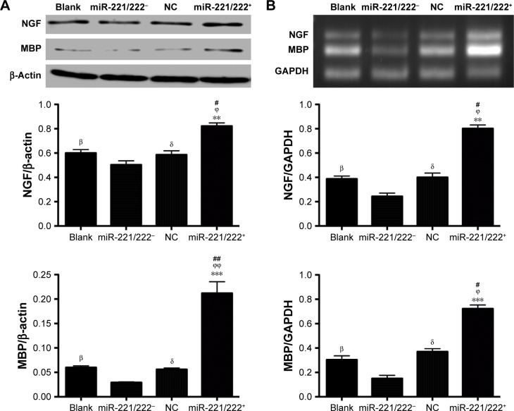
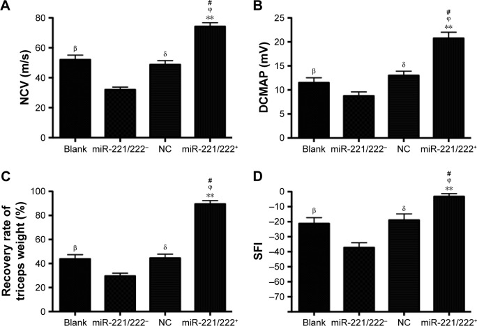
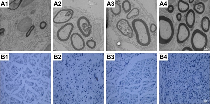
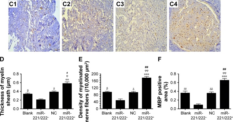
MicroRNA (miRNA) has great potential to treat a wide range of illnesses by regulating the expression of eukaryotic genes. Biomaterials with high transfection efficiency and low toxicity are needed to deliver miRNA to target cells. In this study, a biodegradable and biocompatible cationic polymer (PDAPEI) was synthetized from low molecular weight polyethyleneimine (PEI1.8kDa) cross-linked with 2,6-pyridinedicarboxaldehyde. PDAPEI showed a lower cytotoxicity and higher transfection efficiency than PEI25kDa in transfecting miR-221/222 into rat Schwann cells (SCs). The upregulation of miR-221/222 in SCs promoted the expression of nerve growth factor and myelin basic protein in vitro. The mouse sciatic nerve crush injury model was used to evaluate the effectiveness of PDAPEI/miR-221/222 complexes for nerve regeneration in vivo. The results of electrophysiological tests, functional assessments, and histological and immunohistochemistry analyses demonstrated that PDAPEI/miR-221/222 complexes significantly promoted nerve regeneration after sciatic nerve crush, specifically enhancing remyelination. All these results show that the use of PDAPEI to deliver miR-221/222 may provide a safe therapeutic means of treating nerve crush injury and may help to overcome the barrier of biomaterial toxicity and low efficiency often encountered during medical intervention.

Keywords: PDAPEI; miR-221/222; nerve regeneration; remyelination.


Conflict of interest statement

Disclosure The authors report no conflicts of interest in this work.

Figures

Figure 1
Figure 1
Highly purified SCs are used in this study. Notes: (A) S100-β (red); (B) p75NGFR (green); (C) merged (blue, nucleus). Scale bar, 50 μm. Abbreviation: SCs, Schwann cells.
Figure 2
Figure 2
Synthesis reaction of PDAPEI. Abbreviations: PEI, polyethylenimine; PDAPEI, PEI (1.8 kDa, branched) derivative with 2,6-pyridinedicarboxaldehyde (PDA) linkage.
Figure 3
Figure 3
DNA binding capability of PDAPEI. Notes: (A) Agarose gel electrophoresis of complexes at various N/P ratios. (B) Statistical analysis indicated that the band for miR-221/222 was completely invisible at a N/P ratio of 5 or above for PDAPEI. Data are shown as the mean ± SD (n=3). Abbreviations: PDAPEI, PEI (1.8 kDa, branched) derivative with 2,6-pyridinedicarboxaldehyde (PDA) linkage; PEI, polyethylenimine; SD, standard deviation.
Figure 4
Figure 4
Characteristics of PDAPEI/pDNA complexes. Notes: (A) Particle size, (B) zeta potential. Data are shown as the mean ± SD (n=4) (different N/P). (C1 and C2) Morphology of the complexes was measured using the TEM (scale bar: C1, 100 nm; C2, 0.2 μm). Abbreviations: PDAPEI, PEI (1.8 kDa, branched) derivative with 2,6-pyridinedicarboxaldehyde (PDA) linkage; PEI, polyethylenimine; SD, standard deviation; TEM, transmission electron microscope.
Figure 5
Figure 5
Cytotoxicity to rat SCs in vitro. Notes: Tested with CCK-8 assay (viability cells %) (different N/P). (A) Four-hour posttransfection and (B) 24-h posttransfection. The results have been repeated for three times (blue column, PEI25kDa at the same concentration without miR-221/222; red column, PDAPEI at the same concentration without miR-221/222). Abbreviations: CCK, Cell Counting Kit; PDAPEI, PEI (1.8 kDa, branched) derivative with 2,6-pyridinedicarboxaldehyde (PDA) linkage; PEI, polyethylenimine; SCs, Schwann cells.
Figure 6
Figure 6
Toxicity to main organs and local muscle tissue in vivo. Notes: Measured by frozen section after systemic injection of saline, naked siRNA and complexes at a N/P ratio of 300 (A1C1, the saline control group; A2C2, the naked miR-221/222 group; A3C3, the PDAPEI/miR-221/222 group; A1A3, local right thigh muscle; B1B3, liver; C1C3, kidney. Scale bar, 100 μm; H&E staining, ×200). Abbreviations: H&E, hematoxylin and eosin; PDAPEI, PEI (1.8 kDa, branched) derivative with 2,6-pyridinedicarboxaldehyde (PDA) linkage; PEI, polyethylenimine; si RNA, short interfering RNA.
Figure 7
Figure 7
Transfection efficiency of PDAPEI to SCs. Notes: (A) The naked miR-221/222 group, (B) PEI25kDa/miR-221/222 group (at N/P ratio of 10), (C) PDAPEI/miR-221/222 group (at N/P ratio of 200), (D) The percentage of EGFP-positive cells (%) from different groups, (E) Transfection efficiency of PDAPEI and PEI25kDa at different N/P. Transfection efficiency was measured by flow cytometry (A1C1, observed by light microscopy; A2C2, observed by fluorescence microscopy; Scale bar, 100 μm. *P<0.05, PDAPEI/miR-221/222 group versus PEI25kDa/miR-221/222 group; ϕϕP<0.01, PEI25kDa/miR-221/222 group versus naked miR-221/222 group; ###P<0.001, PDAPEI/miR-221/222 group versus naked miR-221/222 group). Abbreviations: PDAPEI, PEI (1.8 kDa, branched) derivative with 2,6-pyridinedicarboxaldehyde (PDA) linkage; PEI, polyethylenimine; SCs, Schwann cells.
Figure 8
Figure 8
Effect of PDAPEI/miR-221/222 on the expression of nerve growth factor (NGF) and myelin basic protein (MBP) in SCs in vitro. Notes: (A) NGF and MBP expression determined by western-blot analysis and (B) RT-PCR analysis of NGF and MBP miRNA expression. βP<0.05, the blank group versus miR-221/222 group; δP<0.05, the NC group versus miR-221/222 group; #P<0.05, ##P<0.01, miR-221/222+ group versus the blank group; ϕP<0.05, ϕϕP<0.01, miR-221/222+ group versus the NC group; **P<0.01, ***P<0.001, miR-221/222+ group versus miR-221/222 group. Abbreviations: MBP, myelin basic protein; NGF, nerve growth factor; PDAPEI, PEI (1.8 kDa, branched) derivative with 2,6-pyridinedicarboxaldehyde (PDA) linkage; PEI, polyethylenimine; NC, negative control; SCs, Schwann cells.
Figure 9
Figure 9
Functional recovery of rats 4 weeks posttreatment. Notes: (A) NCV, (B) DCMAP, (C) recovery rate of triceps weight, (D) SFI from the blank, miR-221/222, NC, and miR-221/222+ group (n=6 in each group. βP<0.05, the blank group versus miR-221/222 group; δP<0.05, the NC group versus miR-221/222 group; #P<0.05, miR-221/222+ group versus the blank group; ϕP<0.05, miR-221/222+ group versus the NC group; **P<0.01, miR-221/222+ group versus miR-221/222 group). Abbreviations: DCMAP, distal compound motor action potential; NCV, nerve conduction velocity; SFI, sciatic function index; NC, negative control.
Figure 10
Figure 10
Ultrastructure and immunohistochemistry of the regenerated nerve 4 weeks posttreatment. Notes: (A1A4) Histology images by micrographs, scale bar: 2 μm. (B1B4) Histology images stained with methylene blue, scale bar: 50 μm. (C1C4) MBP immunohistochemistry of regenerated nerves. The brown spots indicated MBP-positive area, scale bar: 20 μm (A1C1, miR-221/222 group; A2C2, the blank group; A3C3, NC group; A4C4, miR-221/222+ group). (D) Thickness of myelin sheath. (E) Density of myelinated nerve fibers (/10,000 μm2). (F) MBP-positive area (n=6 in each group. βP<0.05, ββP<0.01 the blank group versus miR-221/222 group; δP<0.05, δδP<0.01 the NC group versus miR-221/222 group; #P<0.05, ##P<0.01 miR-221/222+ group versus the blank group; ϕP<0.05, ϕϕP<0.01, miR-221/222+ group versus the NC group; **P<0.01, ***P<0.001, miR-221/222+ group versus miR-221/222 group). Abbreviations: MBP, myelin basic protein; TEM, transmission electron microscope.
Figure 10
Figure 10
Ultrastructure and immunohistochemistry of the regenerated nerve 4 weeks posttreatment. Notes: (A1A4) Histology images by micrographs, scale bar: 2 μm. (B1B4) Histology images stained with methylene blue, scale bar: 50 μm. (C1C4) MBP immunohistochemistry of regenerated nerves. The brown spots indicated MBP-positive area, scale bar: 20 μm (A1C1, miR-221/222 group; A2C2, the blank group; A3C3, NC group; A4C4, miR-221/222+ group). (D) Thickness of myelin sheath. (E) Density of myelinated nerve fibers (/10,000 μm2). (F) MBP-positive area (n=6 in each group. βP<0.05, ββP<0.01 the blank group versus miR-221/222 group; δP<0.05, δδP<0.01 the NC group versus miR-221/222 group; #P<0.05, ##P<0.01 miR-221/222+ group versus the blank group; ϕP<0.05, ϕϕP<0.01, miR-221/222+ group versus the NC group; **P<0.01, ***P<0.001, miR-221/222+ group versus miR-221/222 group). Abbreviations: MBP, myelin basic protein; TEM, transmission electron microscope.

References

    1. Burnett MG, Zager EL. Pathophysiology of peripheral nerve injury: a brief review. Neurosurgical Focus. 2004;16(5):E1. - PubMed
    1. Li BH, Kim SM, Yoo SB, Kim MJ, Jahng JW, Lee JH. Recombinant human nerve growth factor (rhNGF-β) gene transfer promotes regeneration of crush-injured mental nerve in rats. Oral Surg Oral Med Oral Pathol Oral Radiol. 2012;113(3):e26–e34. - PubMed
    1. Song YS, Joe JH, Joo HW, et al. The effects of granulocyte-colony stimulating factor on regeneration in nerve crush injuries in rats. Neurochem Res. 2016;41(7):1645–1450. - PubMed
    1. Gu X, Ding F, Williams DF. Neural tissue engineering options for peripheral nerve regeneration. Biomaterials. 2014;35(24):6143–6156. - PubMed
    1. Choudhury SR, Hudry E, Maguire CA, Sena-Esteves M, Breakefield XO, Grandi P. Viral vectors for therapy of neurologic diseases. Neuropharmacology. 2016 Feb 21; Epub. - PMC - PubMed

MeSH terms

LinkOut - more resources