

Optic perineuritis: a retrospective case series
- PMID: 28652823
- PMCID: PMC5476656
- DOI: 10.2147/IMCRJ.S125972
Optic perineuritis: a retrospective case series
Abstract
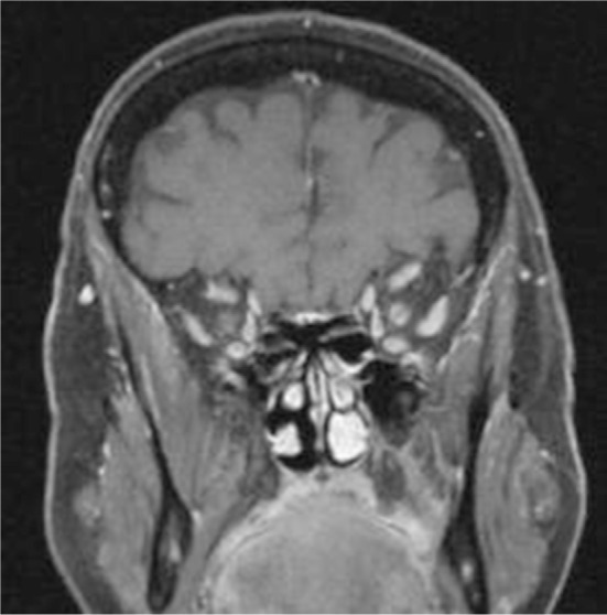
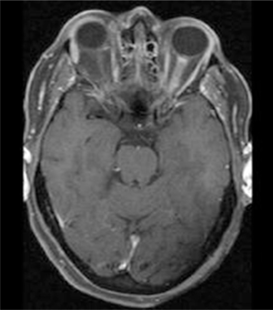
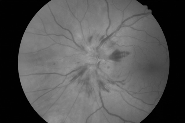
Optic perineuritis (OPN) is a rare inflammatory disorder involving the optic nerve sheath characterized by visual loss. OPN may be isolated and idiopathic or part of an underlying disorder. This case series aimed to help clinicians investigate and manage this disorder. Presentation, clinical findings, and treatment of OPN are discussed. After review of medical records at the ophthalmology clinic at Sahlgrenska University hospital in Gothenburg, Sweden, seven OPN patients (three men and four women) were identified and included in the present case series. These included idiopathic cases and patients with underlying disorders. Age at OPN diagnosis ranged from 26 to 64 years (mean age 55 years, median age 58 years). Five of the patients were treated with corticosteroids. This study suggests that a high-dose course of corticosteroids is important in the treatment of OPN in severely affected patients.
Keywords: corticosteroids; glucocorticoids; idiopathic; inflammation; magnetic resonance imaging; prednisone; vision.
Conflict of interest statement
Disclosure The authors report no conflicts of interest in this work.
Figures
References
-
- Kennerdell JS, Dresner SC. The nonspecific orbital inflammatory syndromes. Surv Ophthalmol. 1984;29(2):93–103. - PubMed
-
- Purvin V, Kawasaki A, Jacobson DM. Optic perineuritis: clinical and radiographic features. Arch Ophthalmol. 2001;119(9):1299–1306. - PubMed
-
- Purvin V, Kawasaki A. Optic perineuritis secondary to Wegener’s granulomatosis. Clin Exp Ophthalmol. 2009;37(7):712–717. - PubMed
-
- Hickman SJ. Optic perineuritis. Curr Neurol Neurosci Rep. 2016;16(2):16. - PubMed
-
- Toshniwal P. Optic perineuritis with secondary syphilis. J Clin Neuroophthalmol. 1987;7(1):6–10. - PubMed
LinkOut - more resources
Full Text Sources
Other Literature Sources
Research Materials
