. 2017 Jun 7;11(3):034116.
doi: 10.1063/1.4985071. eCollection 2017 May.

A magneto-fluidic nanoparticle trapping platform for surface-enhanced Raman spectroscopy

Affiliations

A magneto-fluidic nanoparticle trapping platform for surface-enhanced Raman spectroscopy

Po-Jung Huang et al. Biomicrofluidics. .

Abstract

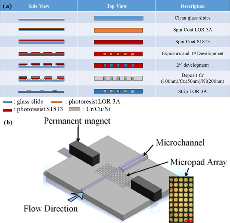
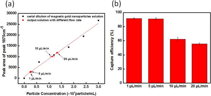
A microfluidic device utilizing magnetically activated nickel (Ni) micropads has been developed for controlled localization of plasmonic core-shell magnetic nanoparticles, specifically for surface enhanced Raman spectroscopy (SERS) applications. Magnetic microfluidics allows for automated washing steps, provides a means for easy reagent packaging, allows for chip reusability, and can even be used to facilitate on-chip mixing and filtration towards full automation of biological sample processing and analysis. Milliliter volumes of gold-coated 175-nm silica encapsulated iron oxide nanoparticles were pumped into a microchannel and allowed to magnetically concentrate down into 7.5 nl volumes over nano-thick lithographically defined Ni micropads. This controlled aggregation of core-shell magnetic nanoparticles by an externally applied magnetic field not only enhances the SERS detection limit within the newly defined nanowells but also generates a more uniform (∼92%) distribution of the SERS signal when compared to random mechanical aggregation. The microfluidic flow rate and the direction and strength of the magnetic field determined the overall capture efficiency of the magneto-fluidic nanoparticle trapping platform. It was found that a 5 μl/min flow rate using an attractive magnetic field provided by 1 × 2 cm neodymium permanent magnets could capture over 90% of the magnetic core-shell nanoparticles across five Ni micropads. It was also observed that the intensity of the SERS signal for this setup was 10-fold higher than any other flow rate and magnetic field configurations tested. The magnetic concentration of the ferric core-shell nanoparticles causes the SERS signal to reach the steady state within 30 min can be reversed by simply removing the chip from the magnet housing and sonicating the retained particles from the outlet channel. Additionally, each magneto-fluidic can be reused without noticeable damage to the micropads up to three times.


Figures

FIG. 1.
FIG. 1.
Magnetically activated Ni-patterned microfluidic device showing (a) fabrication of the micropad array on the platform; (b) schematic diagram of the magnetic platform, composed of a micromagnet array, a microfluidic channel, and a permanent magnet. The inset picture is the micromagnet array. The size of each pad is 50 μm ×50 μm (scale bar is 100 μm).
FIG. 2.
FIG. 2.
Construction of 4-MBA functionalized core-shell magnetic nanoparticles: (a) schematic diagram of the magnetic core-shell nanoparticles, which are composed of (i) an iron core (17 nm), (ii) an SiO2 layer (110 nm), (iii) a gold shell (48 nm), and (iv) a Raman reporter dye molecule (4-MBA); (b) the structure of Raman reporter 4-MBA, (c) TEM images of silica coated iron before gold coating; and (d) SEM images of the core-shell magnetic nanoparticles after coating with gold and functionalization with 4-MBA.
FIG. 3.
FIG. 3.
(a) Brightfield image of micropads aggregating the core-shell magnetic nanoparticles and SERS mapping of the micropads under 10× magnification (scar bar: 50 μm); (b) SEM image of a micropad with magnetically trapped gold coated iron/silica nanoparticles; and (c) comparison of SERS signals of the MBA-functionalized nanoparticle collected from the magnetic micropad area, gap area between pads, and stock solution.
FIG. 4.
FIG. 4.
(a) SERS spectra with attractive, repulsive, and no magnetic fields; and (b) depth and width mapping for SERS signal at 1075 cm−1 with attractive, repulsive, and no magnetic fields.
FIG. 5.
FIG. 5.
Characterization of the solution passing through the magneto-fluidic nanoparticle trapping platform: (a) UV-Vis spectrum of the input and output solutions for the attractive polarity; (b) SERS spectra for core-shell magnetic nanoparticles with different particle concentrations: 3 × 109, 6 × 108, 3 × 107, 1.2 × 107, and 6 × 106 particles/ml; (c) concentration of the standard curve calculated using the data from (b) for core-shell magnetic nanoparticles and a plot of the output solution after magnetic trapping using attractive polarity; and (d) Raman spectra of the output solution for the different polarities: attractive, repulsive, and random (native).
FIG. 6.
FIG. 6.
Characterization of the solution passing through the magneto-fluidic nanoparticle trapping platform using different flow rates. The flow rate used was 1 μl/min, 5 μl/min, 10 μl/min, and 20 μl/min (a) the peak area for the 1075 cm−1 peak for different flow rates as mapped on the serial dilution calibration curve. (b) trapping efficiency of magnetic micropads under different flow rates.
FIG. 7.
FIG. 7.
(a) Schematic diagram for 2D SERS intensity mapping layers. (b) SERS mapping profiles were collected and the average peak area of the normalized of SERS peak at 1075 cm−1 for one Ni micropad at 10, 20, 30, 40, and 50 min. (c) Schematic showing the presumptive aggregation of core-shell magnetic nanoparticles on Ni micropads. (d) Depth mapping of normalized SERS signal at 1075 cm−1 at 10, 20, 30, 40, and 50 min with attractive magnetic fields.
FIG. 8.
FIG. 8.
Recycling and reuse of magneto-fluidic nanoparticle trapping platform. The reuse test is a cyclic process, which consists of three dry-wet cycles. The dry condition is for achieving additional enhancement from magnetic particles on each micropad located in microchannel after water evaporation for static SERS measurements. The wet condition demonstrates trapped magnetic particles concentrated into the 7.5 nl wells over each micropad for dynamic SERS measurement.

References

    1. Huh Y. S., Chung A. J., and Erickson D., Microfluid. Nanofluid. 6, 285–297 (2009).10.1007/s10404-008-0392-3 - DOI
    1. Nie S. and Emory S. R., Science 275, 1102–1106 (1997).10.1126/science.275.5303.1102 - DOI - PubMed
    1. Li M., Cushing S. K., Liang H., Suri S., Ma D., and Wu N., Anal. Chem. 85, 2072–2078 (2013).10.1021/ac303387a - DOI - PubMed
    1. Jackson J. B., Westcott S. L., Hirsch L. R., West J. L., and Halas N. J., Appl. Phys. Lett. 82, 257–259 (2003).10.1103/RevModPhys.57.783 - DOI
    1. Nikoobakht B. and El-Sayed M. A., J. Phys. Chem. A 107, 3372–3378 (2003).10.1021/jp026770+ - DOI

LinkOut - more resources