. 2017 May 26:15:466-482.
doi: 10.1016/j.nicl.2017.05.022. eCollection 2017.

A protocol for manual segmentation of medial temporal lobe subregions in 7 Tesla MRI

Affiliations

A protocol for manual segmentation of medial temporal lobe subregions in 7 Tesla MRI

D Berron et al. Neuroimage Clin. .

Abstract

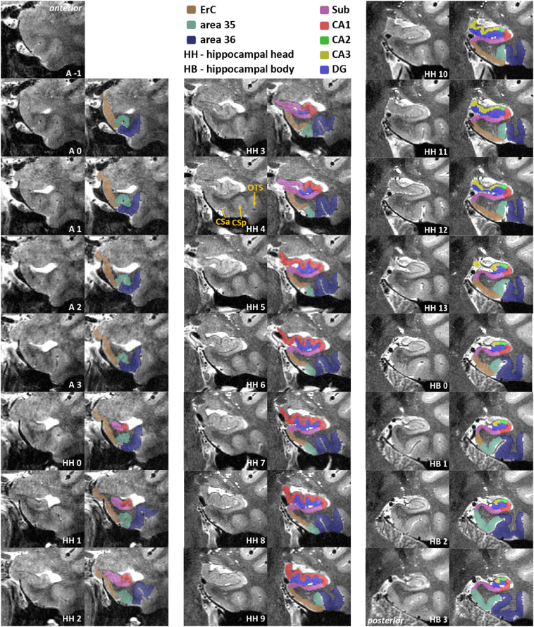
Recent advances in MRI and increasing knowledge on the characterization and anatomical variability of medial temporal lobe (MTL) anatomy have paved the way for more specific subdivisions of the MTL in humans. In addition, recent studies suggest that early changes in many neurodegenerative and neuropsychiatric diseases are better detected in smaller subregions of the MTL rather than with whole structure analyses. Here, we developed a new protocol using 7 Tesla (T) MRI incorporating novel anatomical findings for the manual segmentation of entorhinal cortex (ErC), perirhinal cortex (PrC; divided into area 35 and 36), parahippocampal cortex (PhC), and hippocampus; which includes the subfields subiculum (Sub), CA1, CA2, as well as CA3 and dentate gyrus (DG) which are separated by the endfolial pathway covering most of the long axis of the hippocampus. We provide detailed instructions alongside slice-by-slice segmentations to ease learning for the untrained but also more experienced raters. Twenty-two subjects were scanned (19-32 yrs, mean age = 26 years, 12 females) with a turbo spin echo (TSE) T2-weighted MRI sequence with high-resolution oblique coronal slices oriented orthogonal to the long axis of the hippocampus (in-plane resolution 0.44 × 0.44 mm2) and 1.0 mm slice thickness. The scans were manually delineated by two experienced raters, to assess intra- and inter-rater reliability. The Dice Similarity Index (DSI) was above 0.78 for all regions and the Intraclass Correlation Coefficients (ICC) were between 0.76 to 0.99 both for intra- and inter-rater reliability. In conclusion, this study presents a fine-grained and comprehensive segmentation protocol for MTL structures at 7 T MRI that closely follows recent knowledge from anatomical studies. More specific subdivisions (e.g. area 35 and 36 in PrC, and the separation of DG and CA3) may pave the way for more precise delineations thereby enabling the detection of early volumetric changes in dementia and neuropsychiatric diseases.

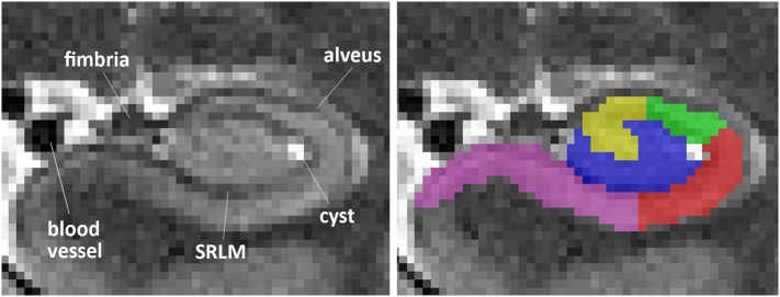
Keywords: AG, Ambient Gyrus; CA1, Cornu Ammonis 1; CA2, Cornu Ammonis 2; CA3, Cornu Ammonis 3; CS, Collateral Sulcus; CSF, Cerebrospinal Fluid; CSa, anterior; CSp, posterior; CaS, Calcarine sulcus; DG, Dentate Gyrus; ErC, Entorhinal Cortex; FG, Fusiform Gyrus; HB, Hippocampal Body; HH, Hippocampal Head; HT, Hippocampal Tail; MTL, Medial Temporal Lobe; OTS, Occipito-temporal Sulcus; PhC, Parahippocampal Cortex; PhG, Parahippocampal Gyrus; PrC, Perirhinal Cortex; SRLM, Stratum radiatum lacunosum-moleculare; SaS, Semiannular Sulcus; Sub, Subiculum.


Figures

Fig. 1
Fig. 1
Segmentation hierarchy. Segmentation of entorhinal cortex (ErC), area 35 and 36 of the perirhinal cortex (PrC), parahippocampal cortex (PhC) and the whole hippocampus separated into head (HH), body (HB) and tail (HT) are described in 2.5.2 (dark blue) and segmentation of hippocampal subfields is described in 2.5.3 (light blue). (For interpretation of the references to colour in this figure legend, the reader is referred to the web version of this article.)
Fig. 2
Fig. 2
Structures excluded from segmentation in a coronal view. Anterior hippocampal body slice from a T2 MRI scan including alveus, fimbria, SRLM, a blood vessel and a cyst in the ventrolateral flexion point of CA1 in the vestigial hippocampal sulcus.
Fig. 3
Fig. 3
Slice-by-slice segmentation for a type 1 collateral sulcus (CS) – anterior part. Slices are 1.1 mm apart. Included are entorhinal cortex (ErC; brown), perirhinal cortex (area 35 in mint green, area 36 in dark blue), subiculum (pink), CA1 (red), CA2 (green), CA3 (yellow) and dentate gyrus (blue). Shown in HH2, ErC covers the ambient gyrus (AG) and superiorly ends at the semiannular sulcus (SaS). SaS constitutes the superior border of ErC and should be extrapolated to anterior slices when it cannot be identified there. (For interpretation of the references to colour in this figure legend, the reader is referred to the web version of this article.)
Fig. 4
Fig. 4
Continuation of Fig. 3 - Slice-by-slice segmentation for a type 1 collateral sulcus (CS) – posterior part. Slices are 1.1 mm apart. Included are parahippocampal cortex (PhC; dark pink), subiculum (pink), CA1 (red), CA2 (green), CA3 (yellow), dentate gyrus (blue), and the hippocampal tail which, is not divided into subfields. In HT7, the subsplenial gyrus starts medially blending into the hippocampus. As soon as it is detached from the hippocampus, it is excluded from segmentation (HT + 1). Delineation of PhC stops at the calcarine sulcus (CaS) in HT2. (For interpretation of the references to colour in this figure legend, the reader is referred to the web version of this article.)
Fig. 5
Fig. 5
Different depths of the collateral sulcus (CS) with respective segmentation rules applied. Sulcus depth is measured from edge to fundus of CS as indicated by the red arrows. Edge, fundus, crown and bank are indicated for easy anatomical descriptions of the gyral and sulcal patterns. Quartiles for segmentation rules are defined by measuring the full extent of grey matter from edge to top along the respective bank as indicated by the white two-sided arrows in the images on the right. Entorhinal cortex (brown) ends laterally at ¼ of the grey matter bank medial to CS, when CS is deeper than 4 mm. For very shallow CS (< 4 mm), entorhinal cortex covers the whole medial bank of CS and ends at the extension of the fundus of CS. Segmentation rules for area 35 (green) change depending on the depth of CS: very deep – area 35 covers the middle part from ¼ to ¾ of the grey matter bank medial to CS; deep – area 35 covers the whole superior ¾ of the grey matter bank medial to CS; shallow - area 35 extends up to half of the lateral bank of CS; very shallow - area 35 extends up to half of the crown of the fusiform gyrus (FG). Area 36 (blue) directly neighbors area 35 laterally, and extends towards the entire bank medial to occipitotemporal sulcus (OTS). The hippocampal head (HH) is depicted in turquoise. (For interpretation of the references to colour in this figure legend, the reader is referred to the web version of this article.)
Fig. 6
Fig. 6
Slice-by-slice segmentation for a type II collateral sulcus (CS) – anterior part. Slices are 1.1 mm apart. Included are entorhinal cortex (ErC; brown), perirhinal cortex (area 35 in mint green, area 36 in dark blue), subiculum (pink), CA1 (red), CA2 (green), CA3 (yellow) and dentate gyrus (blue). HH4 is an example of a transition slice between anterior (CSa) and posterior CS (CSp) and the corresponding segmentation of area 35. The occipitotemporal sulcus (OTS) establishes the lateral border of area 36. (For interpretation of the references to colour in this figure legend, the reader is referred to the web version of this article.)
Fig. 7
Fig. 7
Rules for hippocampal subfield segmentation shown on the relevant slices from anterior to posterior. Schematic descriptions of all rules are depicted in the first column. Specific rule changes or new borders are indicated in red. Dashed lines are used, when the rule in question is inferred from another slice; e.g. the inferior Sub/CA1 border is defined in B, but the superior Sub/CA1 border is defined in B' and extrapolated anteriorly. The relevant anatomical changes are indicated by white labels and arrows in the middle column, e.g. when the uncus separates from the hippocampal body (HB), or the colliculi (Col) disappear. The resulting segmentation is shown in the last column; subiculum (Sub) in pink, CA1 in red, CA2 in green, CA3 in yellow and dentate gyrus (DG) in blue. (For interpretation of the references to colour in this figure legend, the reader is referred to the web version of this article.)
Fig. 8
Fig. 8
Heuristic rules for separation of DG and CA3 if the endfolial pathway is not visible. (1) construct a line from middle most superior part of the hippocampus to medial DG touching Sub; (2) from that point, draw a line laterally along the dark band until Sub starts curving, (3) parallel to this intersect line 1 centrally between SRLM and outermost extent of the hippocampus proper,; (4) centrally intersect line 3 orthogonally; (5) CA3 assigned to voxels superior to lines 3 and 4. Applied rules are shown in the lower panel; unspecific hippocampal body (HB) in turquoise, subiculum (Sub) in pink, CA1 in red, CA2 in green, CA3 in yellow and dentate gyrus (DG) in blue. Compare the right panels for visual segmentation based on the endfolial pathway on the same slice. (For interpretation of the references to colour in this figure legend, the reader is referred to the web version of this article.)
Fig. 9
Fig. 9
Exemplary segmentation profile. This anterior-to-posterior axis (i.e. along the longitudinal axis of the hippocampus) illustrates the key decision points of this protocol (numbers indicate mm distance from the first anterior slice in the protocol). Extrahippocampal regions and hippocampal head (HH), body (HB) and tail (HT) divisions are on the left; hippocampal subfields are depicted on the right. The start and end of each structure are depicted in the same color; often they depend on certain landmarks, e.g. the start and end of HH are used as a reference for the occurrence of the entorhinal cortex (ErC), area 35 and area 36, and the parahippocampal cortex (PhC). Most of these points are variable between brains but usually fall into a similar range as shown here. We recommend identifying these points prior to segmentation.

References

    1. Aly M., Ranganath C., Yonelinas A.P. Detecting changes in scenes: the hippocampus is critical for strength-based perception. Neuron. 2013;78:1127–1137. - PMC - PubMed
    1. Aminoff E.M., Kveraga K., Bar M. The role of the parahippocampal cortex in cognition. Trends Cogn. Sci. 2013;17:379–390. - PMC - PubMed
    1. Apostolova L.G. Relationship between hippocampal atrophy and neuropathology markers: a 7T MRI validation study of the EADC-ADNI harmonized hippocampal segmentation protocol. Alzheimers Dement. 2015;11:139–150. - PMC - PubMed
    1. Baker S., Vieweg P., Gao F., Gilboa A., Wolbers T., Black S.E., Rosenbaum R.S. The human dentate gyrus plays a necessary role in discriminating new memories. Curr. Biol. 2016;26:2629–2634. - PubMed
    1. Bernasconi N., Bernasconi A., Caramanos Z., Antel S.B., Andermann F., Arnold D.L. Mesial temporal damage in temporal lobe epilepsy: a volumetric MRI study of the hippocampus, amygdala and parahippocampal region. Brain. 2003;126:462–469. - PubMed

Publication types