. 2017 Jun 3:15:525-540.
doi: 10.1016/j.nicl.2017.06.005. eCollection 2017.

Neural circuit of verbal humor comprehension in schizophrenia - an fMRI study

Affiliations

Neural circuit of verbal humor comprehension in schizophrenia - an fMRI study

Przemysław Adamczyk et al. Neuroimage Clin. .

Abstract

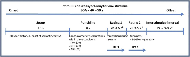
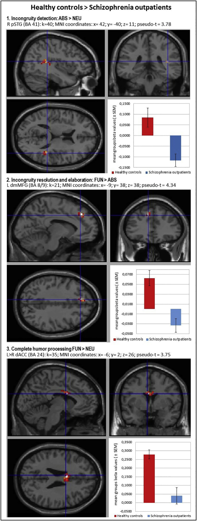
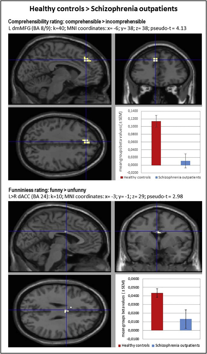
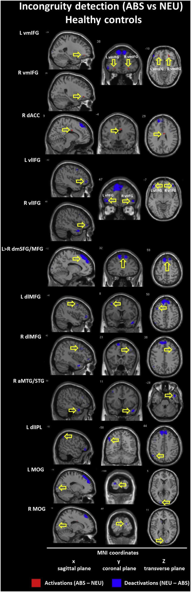
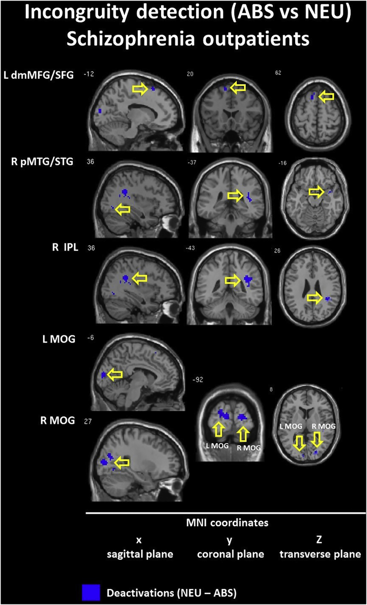
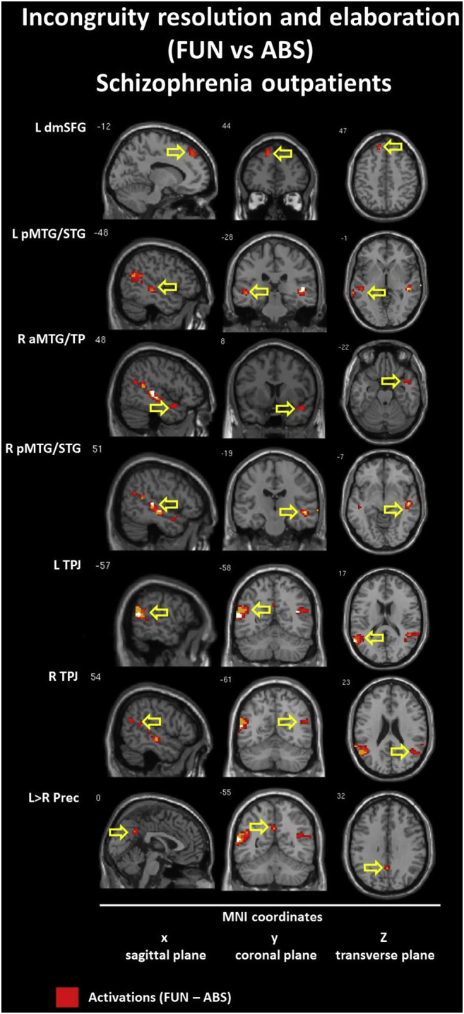
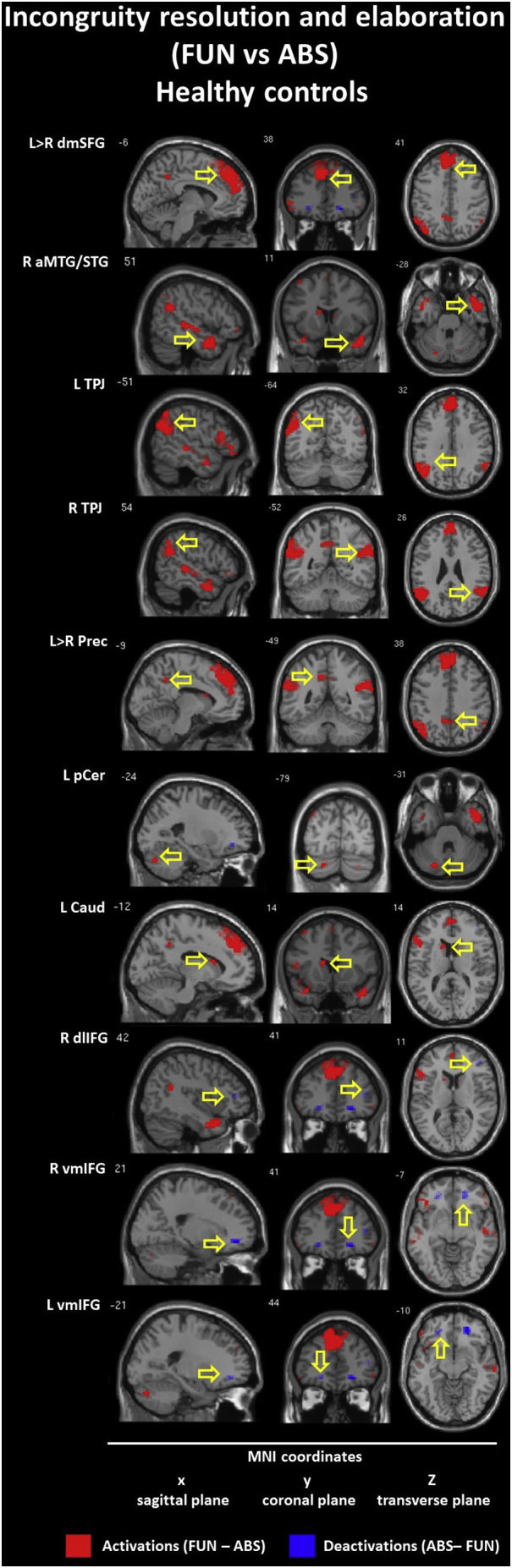
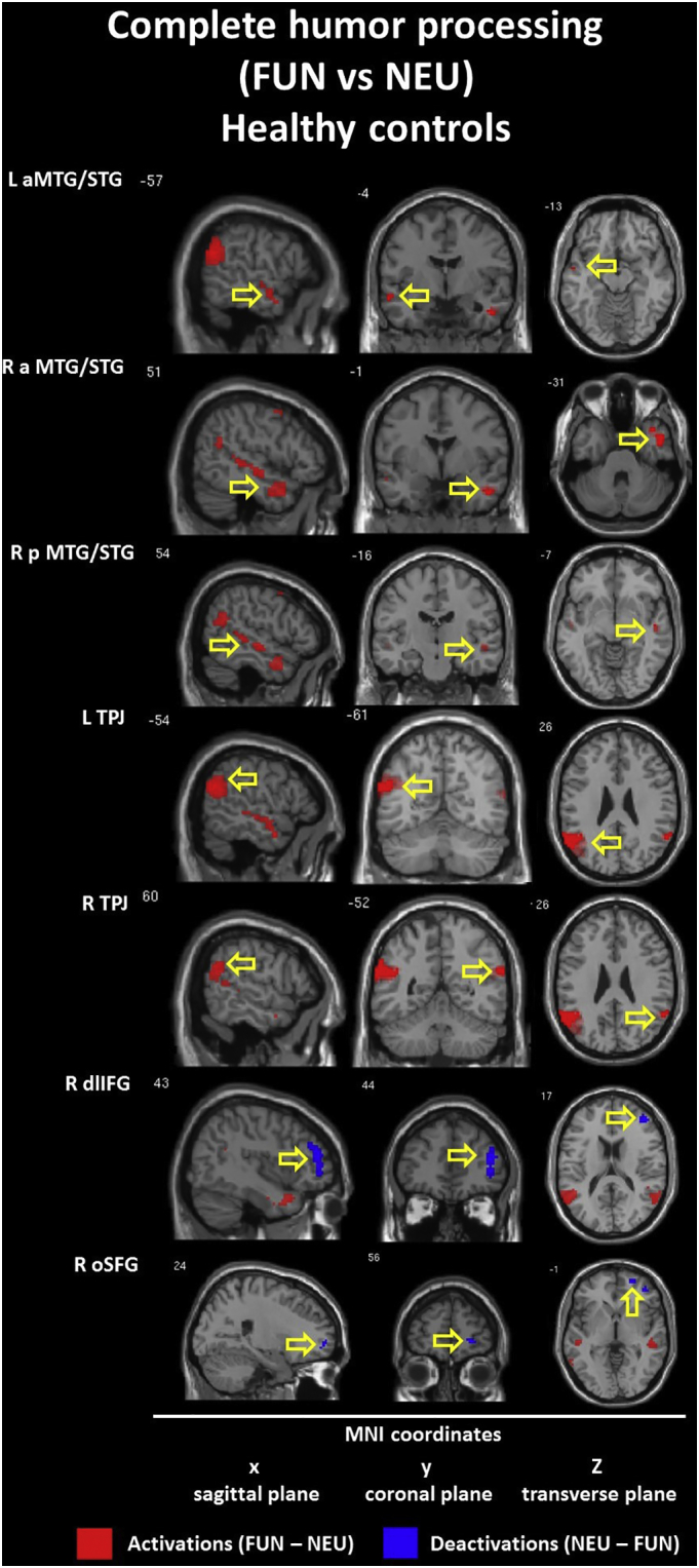
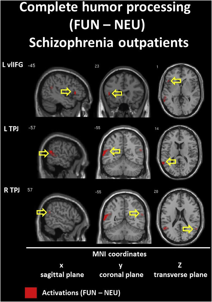
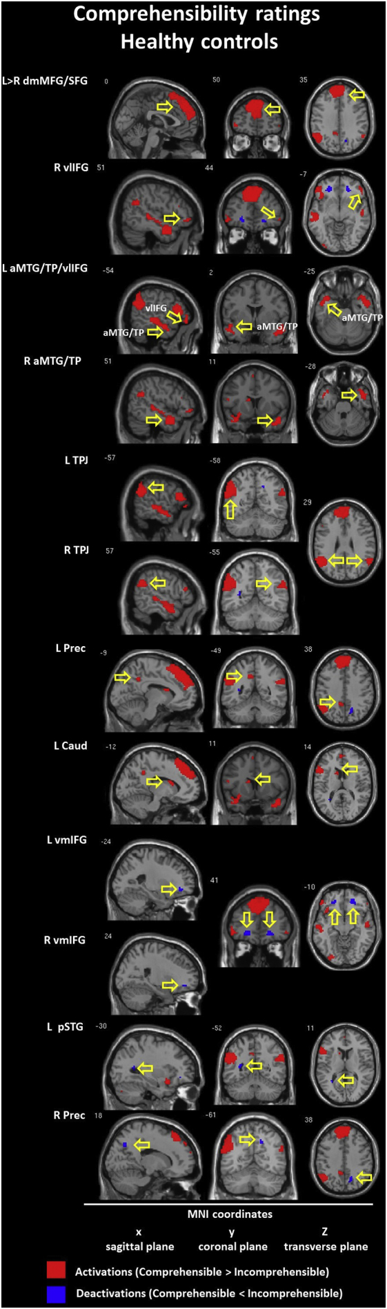
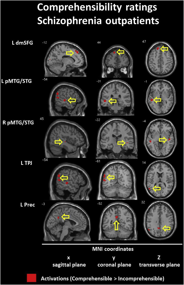
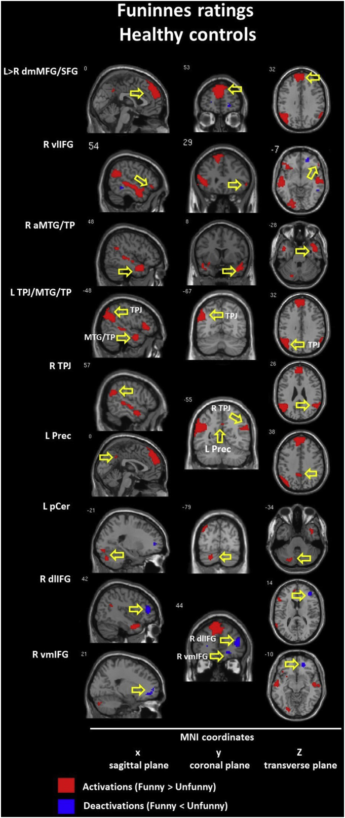
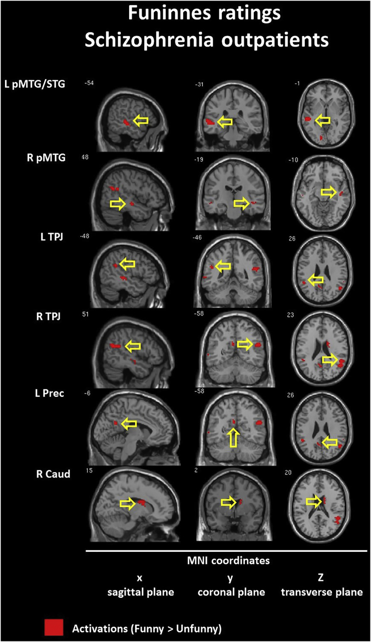
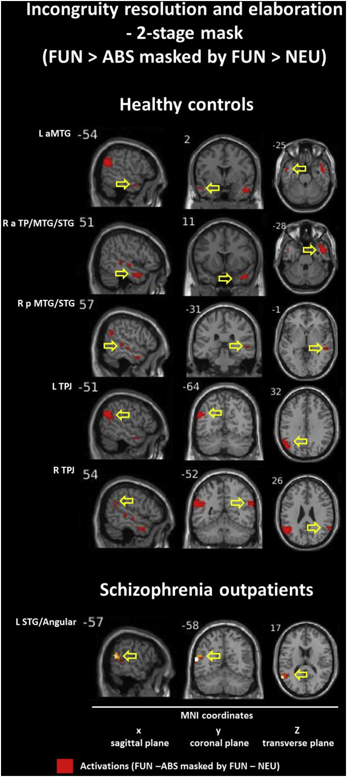
Individuals with schizophrenia exhibit problems with understanding the figurative meaning of language. This study evaluates neural correlates of diminished humor comprehension observed in schizophrenia. The study included chronic schizophrenia (SCH) outpatients (n = 20), and sex, age and education level matched healthy controls (n = 20). The fMRI punchline based humor comprehension task consisted of 60 stories of which 20 had funny, 20 nonsensical and 20 neutral (not funny) punchlines. After the punchlines were presented, the participants were asked to indicate whether the story was comprehensible and how funny it was. Three contrasts were analyzed in both groups reflecting stages of humor processing: abstract vs neutral stories - incongruity detection; funny vs abstract - incongruity resolution and elaboration; and funny vs neutral - complete humor processing. Additionally, parametric modulation analysis was performed using both subjective ratings separately. Between-group comparisons revealed that the SCH subjects had attenuated activation in the right posterior superior temporal gyrus (BA 41) in case of irresolvable incongruity processing of nonsensical puns; in the left dorsomedial middle and superior frontal gyri (BA 8/9) in case of incongruity resolution and elaboration processing of funny puns; and in the interhemispheric dorsal anterior cingulate cortex (BA 24) in case of complete processing of funny puns. Additionally, during comprehensibility ratings the SCH group showed a suppressed activity in the left dorsomedial middle and superior frontal gyri (BA 8/9) and revealed weaker activation during funniness ratings in the left dorsal anterior cingulate cortex (BA 24). Interestingly, these differences in the SCH group were accompanied behaviorally by a protraction of time in both types of rating responses and by indicating funny punchlines less comprehensible. Summarizing, our results indicate neural substrates of humor comprehension processing impairments in schizophrenia, which is accompanied by fronto-temporal hypoactivation.

Keywords: ABS, absurd/nonsensical punchline; ACC, anterior cingulate cortex; BA, Brodmann's area; CON, healthy controls/control group; Communication skills; EEG, electroencephalography; ERPs, EEG event-related potentials; FDR, False Discovery Rate; FUN, funny punchline; FWHM, full-width-at-half-maximum; Figurative meaning; Functional magnetic resonance imaging; GLM, general linear model; Humor; IFG, inferior frontal gyrus; IPL, Inferior Parietal Lobule; ISI, interstimulus-interval; L, left hemisphere; MFG, medial frontal gyrus; MNI, Montreal Neurological Institute coordinates; MOG, middle occipital gyrus; MRI, magnetic resonance imaging; MTG, middle temporal gyrus; MoCA, Montreal Cognitive Assessment; NEU, neutral/unfunny punchline; PANSS, Positive and Negative Syndrome Scale; PFC, prefrontal cortex; R, right hemisphere; RHLB, Right Hemisphere Language Battery; RT, reaction time; SCH, schizophrenia outpatients/clinical group; SD, standard deviations; SEM, standard error of the mean; SFG, Superior Frontal Gyrus; SOA, stimulus onset asynchrony; STG, superior temporal gyrus; Schizophrenia; TP, temporal pole; TPJ, temporoparietal junction; ToM, theory of mind.; dACC, dorsal anterior cingulate cortex; dlPFC, dorsolateral prefrontal cortex; dmMFG, dorsomedial Middle Frontal Gyrus; fMRI, functional magnetic resonance imaging; fNIRS, functional near-infrared spectroscopy; k, number of voxels in analyzed cluster size; ns, non-significant group difference; pSTG, posterior Superior Temporal Gyrus; sLORETA, standardized low resolution brain electromagnetic tomography analysis.


Figures

Fig. 1
Fig. 1
Scheme of experimental stimuli presentation. Abbreviations: # - no time limit for rating responses; * - random order of presentations; ISI – interstimulus-interval; RT - reaction time measurement; SOA – stimulus onset asynchrony; experimental conditions description: ABS – absurd punchline (incomprehensible and unfunny content); FUN – funny punchline (comprehensible and funny content); NEU – neutral punchline (comprehensible and unfunny content).
Fig. 2
Fig. 2
Localizations of brain regions revealed by between-group contrasts during humor comprehension process. Images of the localizations of brain regions activations (upper panels: sagittal and coronal planes; left bottom: transverse plane) were obtained by XjView toolbox (http://www.alivelearn.net/xjview). Each right bottom panel: mean group beta values (± SEM) averaged from the whole analyzed cluster volume presented as arbitrary units. Statistical analysis utilized a non-parametric whole-brain voxel-wise Pseudo-t-test. Localizations are reported as local maximum threshold with k ≥ 10 voxels extent threshold and with False Discovery Rate (FDR) correction at alpha = 0.0500, or uncorrected at alpha = 0.0010. L – left hemisphere; R – right hemisphere; BA – Brodmann's area; k – number of voxels in analyzed cluster size; MNI - Montreal Neurological Institute coordinates; pSTG – posterior Superior Temporal Gyrus; dmMFG – dorsomedial Middle Frontal Gyrus; dACC – dorsal Anterior Cingulate Cortex.
Fig. 3
Fig. 3
Localizations of brain regions revealed by between-group contrasts during comprehensibility and funniness ratings analyzed as parametric modulators. Images of the localizations of brain regions activations (upper panels: sagittal and coronal planes; left bottom: transverse plane) were obtained by XjView toolbox (http://www.alivelearn.net/xjview). Each right bottom panel: mean group beta values (± SEM) averaged from the whole analyzed cluster volume presented as arbitrary units. Statistical analysis utilized a non-parametric whole-brain voxel-wise Pseudo-t-test. Localizations are reported as local maximum threshold with k ≥ 10 voxels extent threshold and with False Discovery Rate (FDR) correction at alpha = 0.0500, or uncorrected at alpha = 0.0010. L – left hemisphere; R – right hemisphere; BA – Brodmann's area; k – number of voxels in analyzed cluster size; MNI - Montreal Neurological Institute coordinates; dmMFG – dorsomedial Middle Frontal Gyrus; dACC – dorsal Anterior Cingulate Cortex.
Fig. 4
Fig. 1
Pattern of brain activations during verbal humor processing - incongruity detection in healthy controls. Fig. 1 Localizations of brain regions revealed by within-group contrasts during incongruity detection in healthy controls (n = 20). Images of the localizations of brain regions activations (left: sagittal; middle: coronal; right: transverse planes) were obtained by XjView toolbox (http://www.alivelearn.net/xjview). Statistical analysis utilized a non-parametric whole-brain voxel-wise Pseudo-t-test. Localizations are reported as local maximum threshold with k ≥ 10 voxels extent threshold and with False Discovery Rate (FDR) correction at alpha = 0.0500, or uncorrected at alpha = 0.0010. L – left hemisphere; R – right hemisphere; MNI - Montreal Neurological Institute coordinates; vmIFG, ventromedial Inferior Frontal Gyrus; dACC, dorsal Anterior Cingulate Cortex; vlIFG, ventrolateral Inferior Frontal Gyrus; dmSFG/MFG, dorsomedial Superior Frontal Gyrus/Middle Frontal Gyrus; dlMFG, dorsolateral Middle Frontal Gyrus; aMTG/STG, anterior Middle Temporal Gyrus/Superior Temporal Gyrus; dlIPL, dorsolateral Inferior Parietal Lobe; MOG, Middle Occipital Gyrus.
Fig. 5
Fig. 2
Pattern of brain activations during verbal humor processing - incongruity detection in schizophrenia outpatients. Fig. 2 Localizations of brain regions revealed by within-group contrasts during incongruity detection in schizophrenia outpatients (n = 20). Images of the localizations of brain regions activations (left: sagittal; middle: coronal; right: transverse planes) were obtained by XjView toolbox (http://www.alivelearn.net/xjview). Statistical analysis utilized a non-parametric whole-brain voxel-wise Pseudo-t-test. Localizations are reported as local maximum threshold with k ≥ 10 voxels extent threshold and with False Discovery Rate (FDR) correction at alpha = 0.0500, or uncorrected at alpha = 0.0010. L – left hemisphere; R – right hemisphere; MNI - Montreal Neurological Institute coordinates; dmMFG/SFG, dorsomedial Middle Frontal Gyrus/Superior Frontal Gyrus; pMTG/STG, posterior Middle Temporal Gyrus/Superior Temporal Gyrus; IPL, Inferior Parietal Lobe; MOG, Middle Occipital Gyrus.
Fig. 6
Fig. 3
Pattern of brain activations during verbal humor processing - incongruity resolution and elaboration in healthy controls. Fig. 3 Localizations of brain regions revealed by within-group contrasts during incongruity resolution and elaboration in healthy controls (n = 20). Images of the localizations of brain regions activations (left: sagittal; middle: coronal; right: transverse planes) were obtained by XjView toolbox (http://www.alivelearn.net/xjview). Statistical analysis utilized a non-parametric whole-brain voxel-wise Pseudo-t-test. Localizations are reported as local maximum threshold with k ≥ 10 voxels extent threshold and with False Discovery Rate (FDR) correction at alpha = 0.0500, or uncorrected at alpha = 0.0010. L – left hemisphere; R – right hemisphere; MNI - Montreal Neurological Institute coordinates; dmSFG, dorsomedial Superior Frontal Gyrus; aMTG/STG, anterior Middle Temporal Gyrus/Superior Temporal Gyrus; TPJ, Temporo-Parietal Junction; Prec, Precuneus; pCer, posterior Cerebellum; Caud, Caudate nucleus; dlIFG, dorsolateral Inferior Frontal Gyrus; vmIFG, ventromedial Inferior Frontal Gyrus.
Fig. 7
Fig. 4
Pattern of brain activations during verbal humor processing - incongruity resolution and elaboration in schizophrenia outpatients. Fig. 4 Localizations of brain regions revealed by within-group contrasts during incongruity resolution and elaboration in schizophrenia outpatients (n = 20). Images of the localizations of brain regions activations (left: sagittal; middle: coronal; right: transverse planes) were obtained by XjView toolbox (http://www.alivelearn.net/xjview). Statistical analysis utilized a non-parametric whole-brain voxel-wise Pseudo-t-test. Localizations are reported as local maximum threshold with k ≥ 10 voxels extent threshold and with False Discovery Rate (FDR) correction at alpha = 0.0500, or uncorrected at alpha = 0.0010. L – left hemisphere; R – right hemisphere; MNI - Montreal Neurological Institute coordinates; dmSFG, dorsomedial Superior Frontal Gyrus; pMTG/STG, posterior Middle Temporal Gyrus/Superior Temporal Gyrus; aMTG/TP, anterior Middle Temporal Gyrus/Temporal Pole; pMTG/STG, posterior Middle Temporal Gyrus/Superior Temporal Gyrus; TPJ, Temporo-Parietal Junction; Prec, Precuneus.
Fig. 8
Fig. 5
Pattern of brain activations during verbal humor processing – complete humor processing in healthy controls. Fig. 5 Localizations of brain regions revealed by within-group contrasts during complete humor processing in healthy controls (n = 20). Images of the localizations of brain regions activations (left: sagittal; middle: coronal; right: transverse planes) were obtained by XjView toolbox (http://www.alivelearn.net/xjview). Statistical analysis utilized a non-parametric whole-brain voxel-wise Pseudo-t-test. Localizations are reported as local maximum threshold with k ≥ 10 voxels extent threshold and with False Discovery Rate (FDR) correction at alpha = 0.0500, or uncorrected at alpha = 0.0010. L – left hemisphere; R – right hemisphere; MNI - Montreal Neurological Institute coordinates; aMTG/STG, anterior Middle Temporal Gyrus/Superior Temporal Gyrus; pMTG/STG, posterior Middle Temporal Gyrus/Superior Temporal Gyrus; TPJ, Temporo-Parietal Junction; dlIFG, dorsolateral Inferior Frontal Gyrus; oSFG, orbital Superior Frontal Gyrus.
Fig. 9
Fig. 6
Pattern of brain activations during verbal humor processing - complete humor processing in schizophrenia outpatients. Fig. 6 Localizations of brain regions revealed by within-group contrasts during complete humor processing in schizophrenia outpatients (n = 20). Images of the localizations of brain regions activations (left: sagittal; middle: coronal; right: transverse planes) were obtained by XjView toolbox (http://www.alivelearn.net/xjview). Statistical analysis utilized a non-parametric whole-brain voxel-wise Pseudo-t-test. Localizations are reported as local maximum threshold with k ≥ 10 voxels extent threshold and with False Discovery Rate (FDR) correction at alpha = 0.0500, or uncorrected at alpha = 0.0010. L – left hemisphere; R – right hemisphere; MNI - Montreal Neurological Institute coordinates; vlIFG, ventrolateral Inferior Frontal Gyrus; TPJ, Temporo-Parietal Junction.
Fig. 10
Fig. 7
Pattern of brain activations during subjective ratings on comprehensibility in healthy controls. Fig. 7 Localizations of brain regions revealed by within-group contrasts during comprehensibility ratings analyzed as parametric modulators in healthy controls (n = 20). Images of the localizations of brain regions activations (left: sagittal; middle: coronal; right: transverse planes) were obtained by XjView toolbox (http://www.alivelearn.net/xjview). Statistical analysis utilized a non-parametric whole-brain voxel-wise Pseudo-t-test. Localizations are reported as local maximum threshold with k ≥ 10 voxels extent threshold and with False Discovery Rate (FDR) correction at alpha = 0.0500, or uncorrected at alpha = 0.0010. L – left hemisphere; R – right hemisphere; MNI - Montreal Neurological Institute coordinates; dmMFG/SFG, dorsomedial Middle Frontal Gyrus/Superior Frontal Gyrus; vlIFG, ventrolateral Inferior Frontal Gyrus; aMTG/TP/vlIFG, anterior Middle Temporal Gyrus/Temporal Pole/Inferior Frontal Gyrus; TPJ, Temporo-Parietal Junction; Prec, Precuneus; Caud, Caudate nucleus; vmIFG, ventromedial Inferior Frontal Gyrus; pSTG, posterior Superior Temporal Gyrus.
Fig. 11
Fig. 8
Pattern of brain activations during subjective ratings on comprehensibility in schizophrenia outpatients. Fig. 8 Localizations of brain regions revealed by within-group contrasts during comprehensibility ratings analyzed as parametric modulators in schizophrenia outpatients (n = 20). Images of the localizations of brain regions activations (left: sagittal; middle: coronal; right: transverse planes) were obtained by XjView toolbox (http://www.alivelearn.net/xjview). Statistical analysis utilized a non-parametric whole-brain voxel-wise Pseudo-t-test. Localizations are reported as local maximum threshold with k ≥ 10 voxels extent threshold and with False Discovery Rate (FDR) correction at alpha = 0.0500, or uncorrected at alpha = 0.0010. L – left hemisphere; R – right hemisphere; MNI - Montreal Neurological Institute coordinates; dmSFG, dorsomedial Superior Frontal Gyrus; pMTG/STG, posterior Middle Temporal Gyrus/Superior Temporal Gyrus; TPJ, Temporo-Parietal Junction; Prec, Precuneus.
Fig. 12
Fig. 9
Pattern of brain activations during subjective ratings on funniness in healthy controls. Fig. 9 Localizations of brain regions revealed by within-group contrasts during funniness ratings analyzed as parametric modulators in healthy controls (n = 20). Images of the localizations of brain regions activations (left: sagittal; middle: coronal; right: transverse planes) were obtained by XjView toolbox (http://www.alivelearn.net/xjview). Statistical analysis utilized a non-parametric whole-brain voxel-wise Pseudo-t-test. Localizations are reported as local maximum threshold with k ≥ 10 voxels extent threshold and with False Discovery Rate (FDR) correction at alpha = 0.0500, or uncorrected at alpha = 0.0010. L – left hemisphere; R – right hemisphere; MNI - Montreal Neurological Institute coordinates; dmMFG/SFG, dorsomedial Middle Frontal Gyrus/Superior Frontal Gyrus; vlIFG, ventrolateral Inferior Frontal Gyrus; aMTG/TP, anterior Middle Temporal Gyrus/Temporal Pole; TPJ, Temporo-Parietal Junction; Prec, Precuneus; pCer, posterior Cerebellum; dlIFG, dorsolateral Inferior Frontal Gyrus; vmIFG, ventromedial Inferior Frontal Gyrus.
Fig. 13
Fig. 10
Pattern of brain activations during subjective ratings on funniness in schizophrenia outpatients. Fig. 10 Localizations of brain regions revealed by within-group contrasts during funniness ratings analyzed as parametric modulators in schizophrenia outpatients (n = 20). Images of the localizations of brain regions activations (left: sagittal; middle: coronal; right: transverse planes) were obtained by XjView toolbox (http://www.alivelearn.net/xjview). Statistical analysis utilized a non-parametric whole-brain voxel-wise Pseudo-t-test. Localizations are reported as local maximum threshold with k ≥ 10 voxels extent threshold and with False Discovery Rate (FDR) correction at alpha = 0.0500, or uncorrected at alpha = 0.0010. L – left hemisphere; R – right hemisphere; MNI - Montreal Neurological Institute coordinates; pMTG/STG, posterior Middle Temporal Gyrus/Superior Temporal Gyrus; TPJ, Temporo-Parietal Junction; Prec, Precuneus; Caud, Caudate nucleus.
Fig. 14
Fig. 11
Pattern of brain activations during verbal humor processing – 2-stage masking for incongruity resolution and elaboration processing in healthy controls and schizophrenia outpatients. Fig. 11 Localizations of brain regions revealed by within-group 2-stage mask contrasts during incongruity resolution and elaboration processing in healthy controls (n = 20) and schizophrenia outpatients (n = 20). Images of the localizations of brain regions activations (left: sagittal; middle: coronal; right: transverse planes) were obtained by XjView toolbox (http://www.alivelearn.net/xjview). Statistical analysis utilized a non-parametric whole-brain voxel-wise Pseudo-t-test. Localizations are reported as local maximum threshold with k ≥ 10 voxels extent threshold and with uncorrected significance level at alpha = 0.0010. L – left hemisphere; R – right hemisphere; MNI - Montreal Neurological Institute coordinates; aTP/MTG/STG, anterior Temporal Pole/Middle Temporal Gyrus/Superior Temporal Gyrus; pMTG/STG, posterior Middle Temporal Gyrus/Superior Temporal Gyrus; TPJ, Temporo-Parietal Junction.

References

    1. Adamczyk P., Daren A., Sułecka A., Błądziński P., Cichocki Ł., Kalisz A., Gawęda Ł., Cechnicki A. Do better communication skills promote sheltered employment in schizophrenia? Schizophr. Res. 2016;176:331–339. - PubMed
    1. Atkins M., Burgess A., Bottomley C., Riccio M. Chlorpromazine equivalents: a consensus of opinion for both clinical and research applications. Psychiatr. Bull. 1997;21:224–226.
    1. Bartolo A., Benuzzi F., Nocetti L., Baraldi P., Nichelli P. Humor comprehension and appreciation: an FMRI study. J. Cogn. Neurosci. 2006;18:1789–1798. - PubMed
    1. Bekinschtein T.A., Davis M.H., Rodd J.M., Owen A.M. Why clowns taste funny: the relationship between humor and semantic ambiguity. J. Neurosci. 2011;31:9665–9671. - PMC - PubMed
    1. Benton E., Bryan K. Right cerebral hemisphere damage: incidence of language problems. Int. J. Rehabil. Res. Int. Z. Rehabil. Rev. Int. Rech. Readaptation. 1996;19:47–54. - PubMed

Publication types