. 2017 May 29:9:10-20.
doi: 10.1016/j.dadm.2017.05.005. eCollection 2017.

Assessment of amyloid β in pathologically confirmed frontotemporal dementia syndromes

Affiliations

Assessment of amyloid β in pathologically confirmed frontotemporal dementia syndromes

Rachel H Tan et al. Alzheimers Dement (Amst). .

Abstract

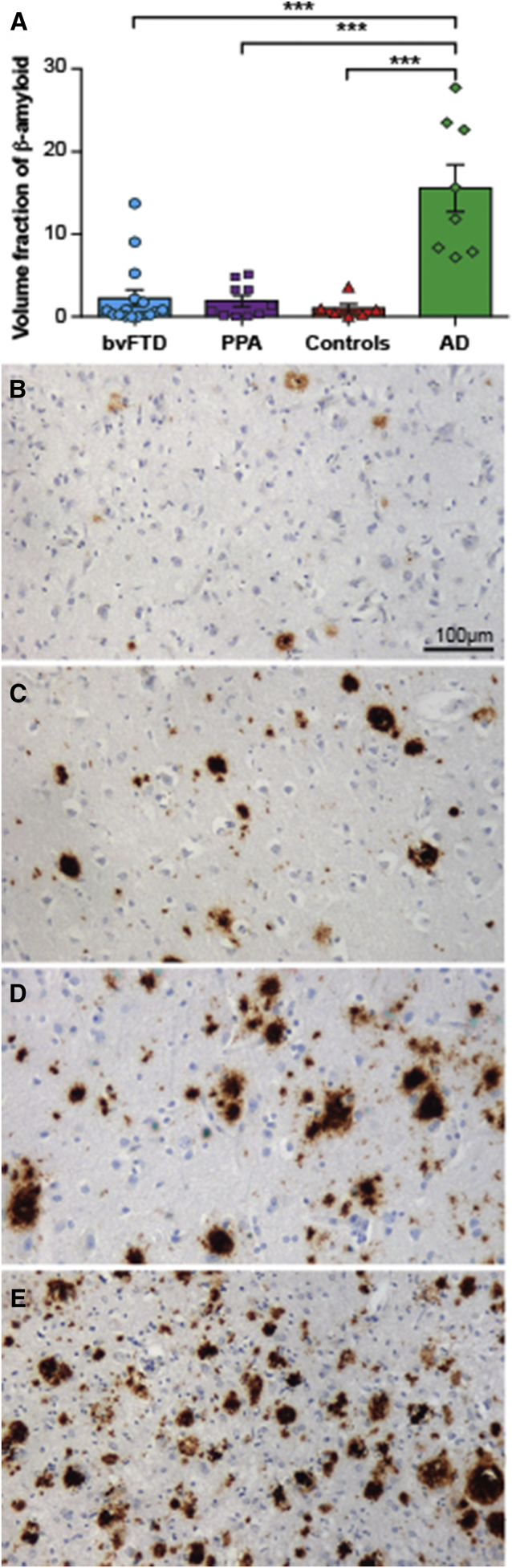
Introduction: The diagnostic utility of in vivo amyloid β (Aβ) imaging to aid in the clinical distinction between frontotemporal dementia (FTD) and Alzheimer's disease remains unclear without data on the prevalence and severity of Aβ in pathologically confirmed FTD syndromes.

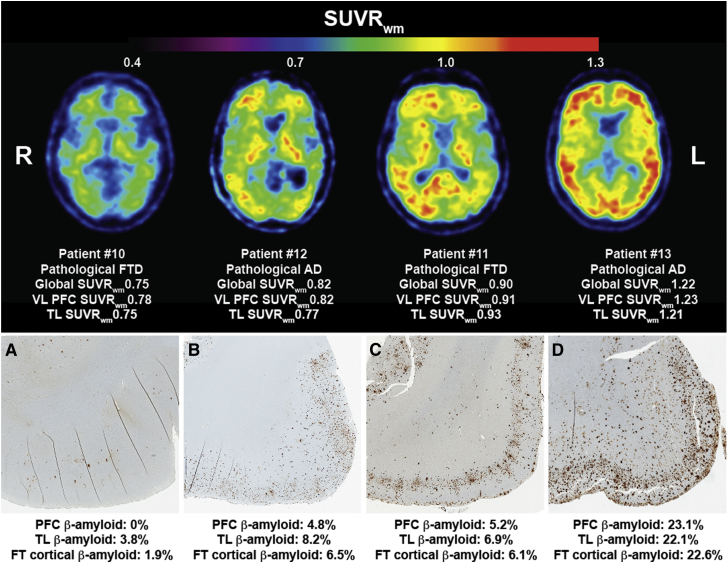
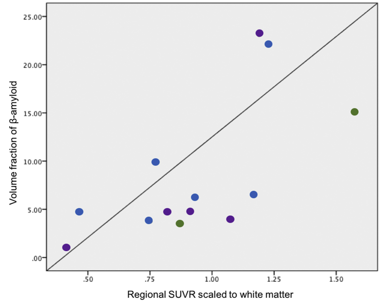
Methods: Aβ was assessed in 98 autopsy-confirmed FTD and 36 control cases, and the pathological accuracy of 11C-Pittsburgh compound B (PiB)-positron emission tomography imaging was assessed in a subset of FTD cases (n = 15).

Results: Aβ was identified in a similar proportion of FTD syndromes and age-matched controls and increases with age. Alzheimer's disease pathology was identified in all cases with high PiB retention and in one case with low PiB retention. We further demonstrate a strong regional correlation between volume fraction of histological Aβ with PiB standard uptake value ratio scaled to the white matter.

Discussion: The present study provides a pathologic reference to assist in the interpretation of in vivo assessments in FTD syndromes.

Keywords: 11C-Pittsburgh compound B; Alzheimer's disease; Amyloid β; Diagnostic; Frontotemporal dementia syndromes.


Figures

Fig. 1
Fig. 1
The proportion of Aβ positivity and topographical distribution of Aβ in frontotemporal dementia (FTD) syndromes and age-matched controls. The proportion of patients that demonstrate no Aβ deposition (A0), Aβ in the frontal-temporal cortices (A1), additional Aβ in the basal ganglia (A2), and additional Aβ in the substantia nigra (A3) is shown in patients with behavioral variant frontotemporal dementia (bvFTD), semantic variant primary progressive aphasia (sv-PPA), non-fluent variant primary progressive aphasia (nfv-PPA), and age-matched controls. Table inset demonstrates the percentage of cases with any Aβ deposition by age. As only one nfv-PPA and one control demonstrated an A3 distribution, these have been combined with the A2 group in the present figure.
Fig. 2
Fig. 2
The proportion of Aβ positivity and topographical distribution of Aβ in frontotemporal dementia (FTD) syndromes with and without motor impairment. The proportion of patients that demonstrate no Aβ deposition (A0), Aβ in the frontal-temporal cortices (A1), additional Aβ in the basal ganglia (A2), and additional Aβ in the substantia nigra (A3) is shown in patients with behavioral variant FTD cases without motor impairment (bvFTD), primary progressive aphasia cases without motor impairment (PPA), cases with motor neuron disease (MND), and cases with extrapyramidalism. As only one PPA case without motor impairment had an A3 distribution (4%, 1/26), this has been combined with the A2 group.
Fig. 3
Fig. 3
The proportion of Aβ positivity and topographical distribution of Aβ in frontotemporal dementia (FTD) cases with and without a genetic mutation. The proportion of patients that demonstrate no Aβ deposition (A0), Aβ in the frontal-temporal cortices (A1), additional Aβ in the basal ganglia (A2), and additional Aβ in the substantia nigra (A3) is shown in FTD cases without a mutation (n = 55), a MAPT mutation (n = 4), a C9ORF72 expansion (n = 10), and a GRN mutation (n = 7) is shown. As only one FTD case without a mutation had an A3 distribution (1%, 1/73), this has been combined with the A2 group.
Fig. 4
Fig. 4
PiB-PET SUVRwm and cortical Aβ pathology in patients with clinical FTD. A strong regional correlation was identified between the volume fraction of histological Aβ deposited in the frontal and temporal cortices with the corresponding SUVRwm in patients with clinical FTD (P <.05). Abbreviations: FT, frontotemporal; FTD, frontotemporal dementia; PET, positron emission tomography; PiB, 11C-Pittsburgh compound B; SUVRwm, standard uptake value ratio scaled to the white matter; TL, temporal lobe; VL PFC, ventrolateral prefrontal cortex.
Fig. 5
Fig. 5
Volume fraction of cortical Aβ in frontotemporal dementia (FTD), Alzheimer's disease (AD), and controls ≤75 years at death. Graph demonstrating the mean (±standard error) volume fraction of histological Aβ in the frontal and temporal cortices of behavioral variant frontotemporal dementia (bvFTD), primary progressive aphasia (PPA), controls, and AD (A). The Aβ observed in the frontal cortex of two sporadic FTD cases (B, C); an FTD case with a C9ORF72 expansion (D); and an AD case (E). ***P < .001.
Supplementary Figure 1

References

    1. Chare L., Hodges J.R., Leyton C.E., McGinley C., Tan R.H., Kril J.J. New criteria for frontotemporal dementia syndromes: clinical and pathological diagnostic implications. J Neurol Neurosurg Psychiatry. 2014;85:865–870. - PubMed
    1. Shelley B.P., Hodges J.R., Kipps C.M., Xuereb J.H., Bak T.H. Is the Pathology of Corticobasal Syndrome Predictable in Life? Movement Disord. 2009;24:1593–1599. - PubMed
    1. Dubois B., Feldman H.H., Jacova C., Hampel H., Molinuevo J.L., Blennow K. Advancing research diagnostic criteria for Alzheimer's disease: the IWG-2 criteria. Lancet Neurol. 2014;13:614–629. - PubMed
    1. Ikonomovic M.D., Klunk W.E., Abrahamson E.E., Mathis C.A., Price J.C., Tsopelas N.D. Post-mortem correlates of in vivo PiB-PET amyloid imaging in a typical case of Alzheimer's disease. Brain. 2008;131:1630–1645. - PMC - PubMed
    1. Lockhart A., Lamb J.R., Osredkar T., Sue L.I., Joyce J.N., Ye L. PIB is a non-specific imaging marker of amyloid-beta (A beta) peptide-related cerebral amyloidosis. Brain. 2007;130:2607–2615. - PubMed