Review
. 2017 Jul 21;15(27):5656-5668.
doi: 10.1039/c7ob01058c. Epub 2017 Jun 27.

Targeting heparin and heparan sulfate protein interactions

Affiliations
Review

Targeting heparin and heparan sulfate protein interactions

Ryan J Weiss et al. Org Biomol Chem. .

Abstract

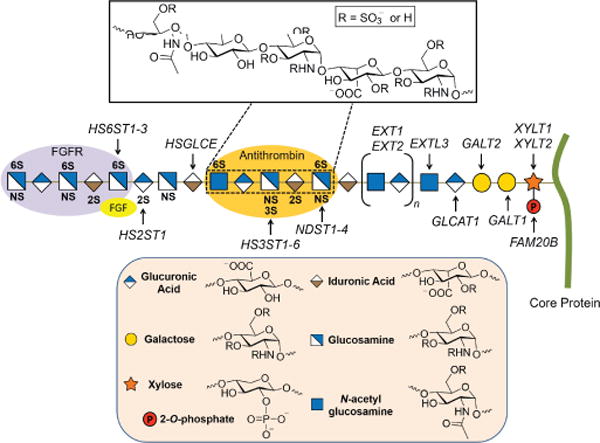
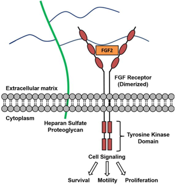
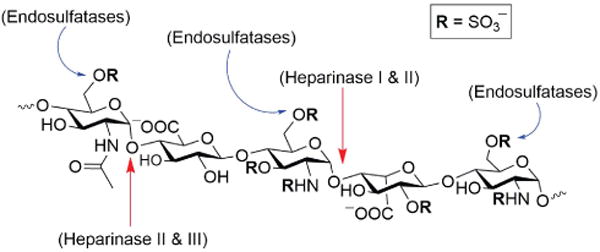
Heparin and heparan sulfate glycosaminoglycans are long, linear polysaccharides that are made up of alternating dissacharide sequences of sulfated uronic acid and amino sugars. Unlike heparin, which is only found in mast cells, heparan sulfate is ubiquitously expressed on the cell surface and in the extracellular matrix of all animal cells. These negatively-charged glycans play essential roles in important cellular functions such as cell growth, adhesion, angiogenesis, and blood coagulation. These biomolecules are also involved in pathophysiological conditions such as pathogen infection and human disease. This review discusses past and current methods for targeting these complex biomolecules as a novel therapeutic strategy to treating disorders such as cancer, neurodegenerative diseases, and infection.


Figures

Figure 1
Figure 1
Heparan sulfate structure and biosynthesis. Respective O- and N-sulfotransferases (HS2ST, HS3ST, HS6ST, and NDST) sulfate specific sites along the HS chain (shown in bold). The purple and orange oval shapes depict protein binding sites along the HS chain. Inset: Pentasaccharide antithrombin (AT) binding sequence.
Figure 2
Figure 2
FGF2 binds to HS (blue lines) and forms a ternary complex with its receptor.
Figure 3
Figure 3
Heparinase (I-III) and endosulfatase (Sulf1, Sulf2) activity on a pentasaccharide region of heparin/HS.
Figure 4
Figure 4
Examples of GAG mimetic compounds.
Figure 5
Figure 5
Examples of cationic antagonists of heparin and HS.
Figure 6
Figure 6
Synthetic self-assembling ligands (11) and UHRA (12) for multivalent binding to heparin. Adapted from Ref. with permission from the Royal Society of Chemistry.
Figure 7
Figure 7
Small molecule antagonists of heparin and HS-protein interactions.
Figure 8
Figure 8
Foldamers: small molecule peptide mimetics for neutralizing heparin.
Figure 9
Figure 9
Structures of surfen (20) and its analogs. Modifications are highlighted in red and green. Reproduced from Ref. with permission from the Royal Society of Chemistry.

References

    1. Bishop JR, Schuksz M, Esko JD. Nature. 2007;446:1030–1037. - PubMed
    1. Esko JD, Lindahl U. J Clin Invest. 2001;108:169–173. - PMC - PubMed
    1. Dhoot GK, Gustafsson MK, Ai X, Sun W, Standiford DM, Emerson CP., Jr Science. 2001;293:1663–1666. - PubMed
    1. Sarrazin S, Lamanna WC, Esko JD. Csh Perspect Biol. 2011;3 - PMC - PubMed
    2. Varki A. Essentials of glycobiology. 2nd. Cold Spring Harbor Laboratory Press; Cold Spring Harbor, N.Y.: 2009. - PubMed
    1. Xu D, Esko JD. Annual Review of Biochemistry. 2014;83:129–157. - PMC - PubMed

MeSH terms