

In Vivo and Mechanistic Studies on Antitumor Lead 7-Methoxy-4-(2-methylquinazolin-4-yl)-3,4-dihydroquinoxalin-2(1H)-one and Its Modification as a Novel Class of Tubulin-Binding Tumor-Vascular Disrupting Agents
- PMID: 28653846
- PMCID: PMC6432920
- DOI: 10.1021/acs.jmedchem.7b00273
In Vivo and Mechanistic Studies on Antitumor Lead 7-Methoxy-4-(2-methylquinazolin-4-yl)-3,4-dihydroquinoxalin-2(1H)-one and Its Modification as a Novel Class of Tubulin-Binding Tumor-Vascular Disrupting Agents
Abstract
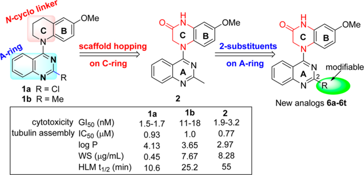
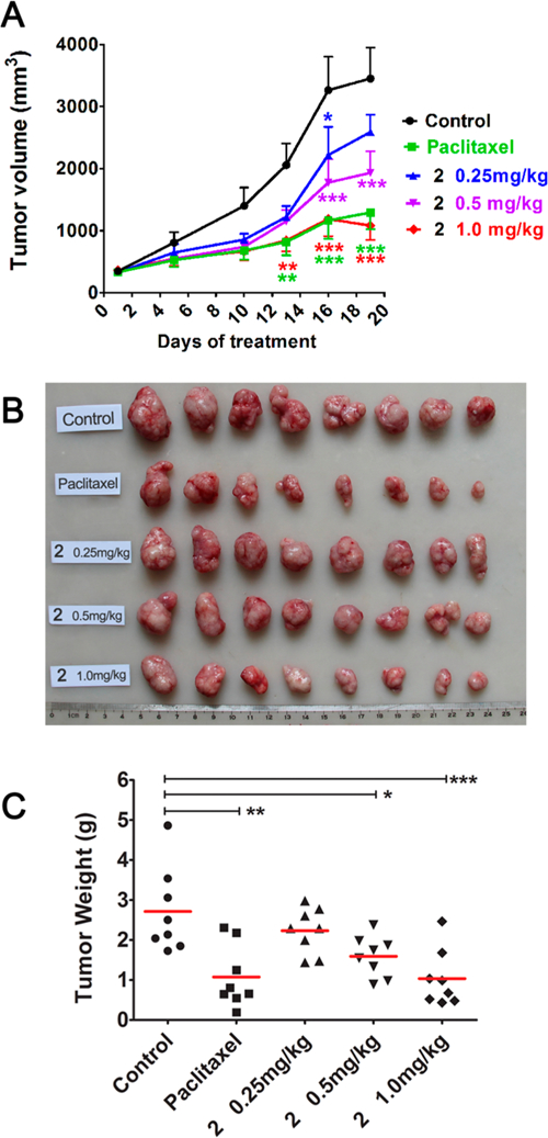
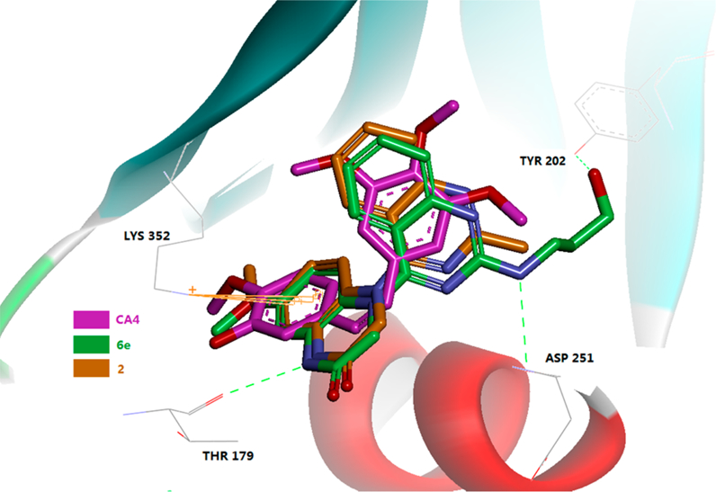
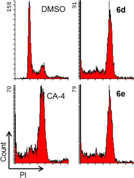
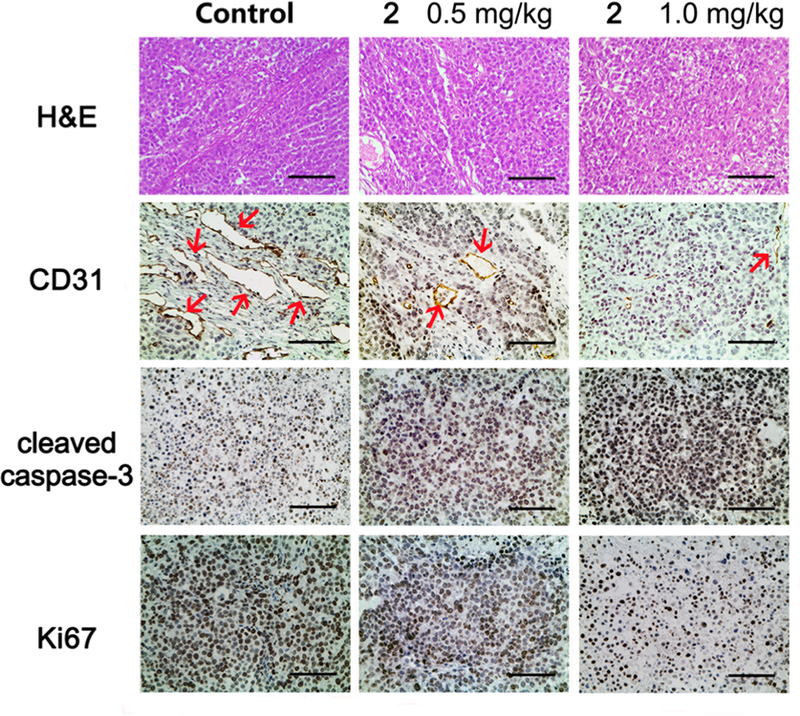
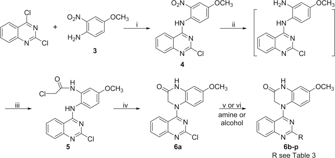
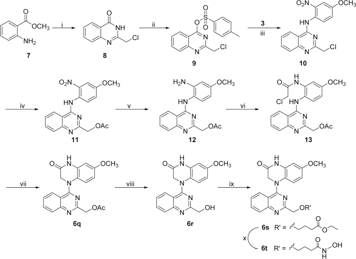
7-Methoxy-4-(2-methylquinazolin-4-yl)-3,4-dihydroquinoxalin-2(1H)-one (2), a promising anticancer lead previously identified by us, inhibited tumor growth by 62% in mice at 1.0 mg/kg without obvious signs of toxicity. Moreover, compound 2 exhibited extremely high antiproliferative activity in the NIH-NCI 60 human tumor cell line panel, with low to sub-nanomolar GI50 values (10-10 M level). It also showed a suitable balance between aqueous solubility and lipophilicity, as well as moderate metabolic stability in vivo. Mechanistic studies using Mayer's hematoxylin and eosin and immunohistochemistry protocols on xenograft tumor tissues showed that 2 inhibited tumor cell proliferation, induced apoptosis, and disrupted tumor vasculature. Moreover, evaluation of new synthetic analogues (6a-6t) of 2 indicated that appropriate 2-substitution on the quinazoline ring could enhance antitumor activity and improve druglike properties. Compound 2 and its analogues with a 4-(2-methylquinazolin-4-yl)-3,4-dihydroquinoxalin-2(1H)-one scaffold thus represent a novel class of tubulin-binding tumor-vascular disrupting agents (tumor-VDAs) that target established blood vessels in tumors.
Conflict of interest statement
The authors declare no competing financial interest.
Figures

References
-
- Perez-Perez MJ; Priego EM; Bueno O; Martins MS; Canela MD; Liekens S Blocking Blood Flow to Solid Tumors by Destabilizing Tubulin: an Approach to Targeting Tumor Growth. J. Med. Chem 2016, 59, 8685–8711. - PubMed
-
- Kaur R; Kaur G; Gill RK; Soni R; Bariwal J Recent Developments in Tubulin Polymerization Inhibitors: an Overview. Eur. J. Med. Chem 2014, 87, 89–124. - PubMed
-
- Tozer GM; Kanthou C; Baguley BC Disrupting Tumor Blood Vessels. Nat. Rev. Cancer 2005, 5, 423–435. - PubMed
Publication types
MeSH terms
Substances
Grants and funding
LinkOut - more resources
Full Text Sources
Other Literature Sources
